

阿尔茨海默病(Alzheimer's disease, AD)是全球最常见的神经退行性疾病,也是导致老年人残疾和依赖的主要原因。阿尔茨海默病被认为是一种人类特有的疾病,尽管其他一些动物也有类似阿尔茨海默病的病理表现。齿动物(齿鲸)与人类有共同的特征,这表明它们可能易患AD。我们用免疫组织化学的方法检测了5种不同物种的22只搁浅齿状突的大脑,以调查是否存在AD的神经病理学特征:淀粉样蛋白- β斑块,磷酸化tau蛋白积聚和神经胶质增生。免疫组化显示所有老龄动物均有淀粉样斑块病理。在3种不同种类的牙突动物中,淀粉样蛋白- β斑块、过度磷酸化tau蛋白的神经元内聚集、神经绒毛线和神经炎斑块共存。1只动物出现发育良好的神经纤毛线、磷酸化tau蛋白聚集和神经炎斑块,但无淀粉样斑块。小胶质细胞和星形胶质细胞与预期一样存在于所有检测的脑样本中,但研究人员观察到细胞形态和数量在个体动物之间存在差异。同时出现的淀粉样β斑块和过度磷酸化的tau蛋白病理在齿突动物的大脑中表明,这三个物种自发地发展出AD样神经病理。
这种病理对动物健康和最终死亡的意义仍有待确定。然而,它可能是导致某些齿目物种无法解释的活体搁浅的原因,并支持“患病领袖”理论,即健康的同类由于高度的社会凝聚力而集体搁浅。

FIG1
Immunolabelling (brown pigment) of amyloid-beta peptide (Aβ) and amyloid plaques (APs) in the cerebrocortical grey matter of odontocetes. Aβ was present in the cell bodies of large neurons within the cerebrocortical grey matter and the labelling frequently extended into the axons (a, supralimbic/paralimbic/limbic lobes, animal Gg2). Intense labelling of Aβ was present in and around the nuclei in a small number of neurons with intensely labelled cytoplasm (b, limbic and anterior paralimbic lobes, animal Gm1). Some odontocetes had minimal intraneuronal labelling of Aβ represented by fine granular deposits within the cytoplasm (c, animal La1). Labelling of Aβ was present in and around a small number of the larger blood vessel (d, limbic and anterior paralimbic lobes, animal La5). When present, large deposits of Aβ in the neuropil in cerebrocortical layer I were diffuse, irregularly shaped with poorly defined borders with some coalescence forming APs present (e, supralimbic/paralimbic/limbic lobes, animal Tt1). When abundant, APs frequently coalesced (f, supralimbic/paralimbic/limbic lobes, animal Gm1; g, supralimbic/paralimbic/limbic lobes, animal Tt1). APs were distributed sparsely in animal La5 and did not coalesce (h, limbic and anterior paralimbic lobes). APs varied in shape and size within the same animal (i, limbic and anterior paralimbic lobes; j, supralimbic/paralimbic/limbic lobes; k, lingual lobe, all animal La5). Animal Gm1 had a medium number of APs present in the cerebrocortical grey matter, some of which had coalesced (l, supralimbic/paralimbic/limbic lobes). Animal Tt1 had the largest number of APs present, and these extended throughout all sections of cerebrocortical grey matter examined (m, supralimbic/paralimbic/limbic lobe). Positive control sections were composed of human brain tissue from a case of definitely diagnosed Alzheimer's disease (n). Sections used for negative controls were composed of semi-serial sections of human brain tissue from a case of definitely diagnosed Alzheimer's disease (o) or positive odontocete brain tissue and were devoid of any immunolabelling
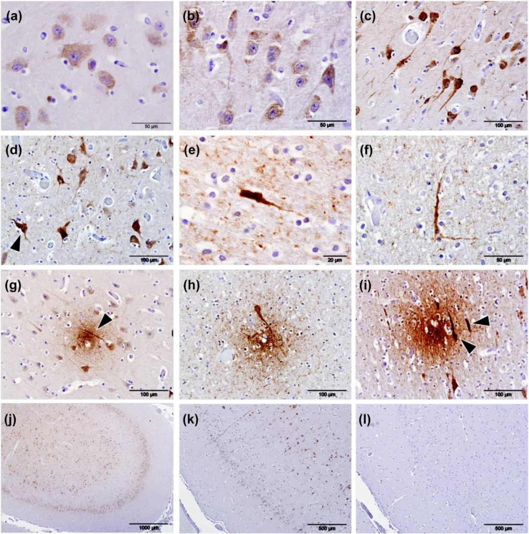
FIG2
Immunolabelling (brown pigment) of phosphorylated tau protein (pTau) using antibody AT180 in the cerebral cortices of odontocetes. When at lower intensities, immunolabelling of pTau was present as cytoplasmic granules within the cell bodies of neurons (a), and with increased intensity of labelling, pTau frequently extended into the axons and dendrites (b,c). Irrespective of the intensity of cytoplasmic labelling, nuclei were consistently devoid of any labelling (c) although neurons that labelled intensely for pTau frequently contained pyknotic nuclei (d, arrowhead). Intensely phospho-tau-stained cells were found in animal (Gm5) and present in all sections of cerebral cortex available for examination (e,f). Neuritic plaques were present in both the white and grey matter in all four aged animals examined (Gm1, Gm5, La5 and Tt1) and within the grey matter often colocalized with neuropil threads (g, arrowhead). Similarly, neuritic plaques colocalized with phospho-tau-positive cell bodies (h,i, arrowheads). pTau labelling was present predominantly in cerebrocortical layers II and V (j). Positive control sections were composed of human brain tissue from a case of definitely diagnosed Alzheimer's disease (k). Semi-serial sections were used for the controls (k, positive; l, negative control)
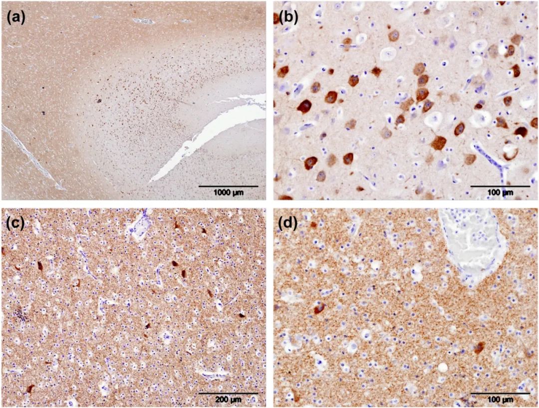
FIG3
Learning techniques used in Knowledge schoolsPhosphorylated tau (pTau) localized using antibody AT180 (brown pigment), in a young bottlenose dolphin (Tt2). pTau was present within neuropil of the white matter but not the grey matter (a). It was also present in the cytoplasm of nearly all neurons in the cerebrocortical grey matter but absent from the nuclei and surrounding neuropil (b). Furthermore, it was present within the neuropil of the white matter of the supralimbic/paralimbic/limbic lobes of the cortex and the cytoplasm of neurons contained within it (c). Greater magnification of cerebrocortical white matter highlighted the intense cytoplasmic labelling of neurons for pTau while sparing their nuclei (d)
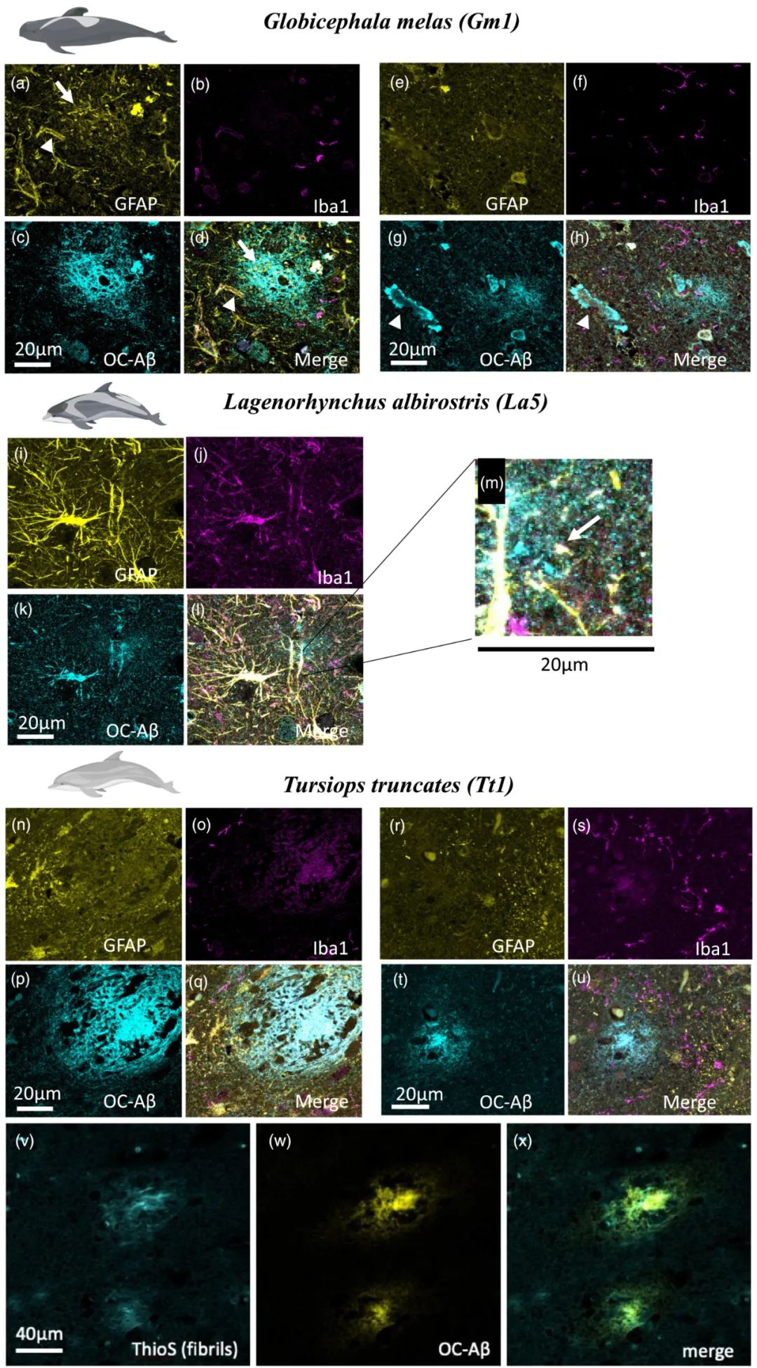
FIG4
GFAP (yellow), Iba1 (magenta) and Aβ fibril (cyan) immunofluorescence labelling in Globicephala melas (Gm1) (a–h), Lagenorhynchus albirostris (La5) (i–m) and Tursiops truncatus (Tt1) (n–u). Gm1 shows diffuse plaques (d,h), evidence of CAA (g, arrowhead) and localized presence of astrocyte processes near some (a, arrow) but not all (e) plaques. Microglia are located near to, but not within plaques (b,f). La5 shows extremely strong GFAP labelling (i), which bled through to other channels. Small diffuse plaques were present, including next to blood vessels (zoomed in section part label m, arrow) but no strong evidence of plaque-associated gliosis. Tt1 showed more punctate astrocyte labelling (n,r) with little association with plaques despite the presence of many diffuse (u) and the occasional dense-core (q) plaques. Although there was no association of microglia with the dense-core plaque (o), there were some microglial processes near the edge of a diffuse plaque (s). We confirmed dense-core plaques contained fibrils using Thioflavin S staining (v), which appeared in amyloid plaques stained for Aβ (w,x). Scale bars = 20 or 40 μm as indicated on panels
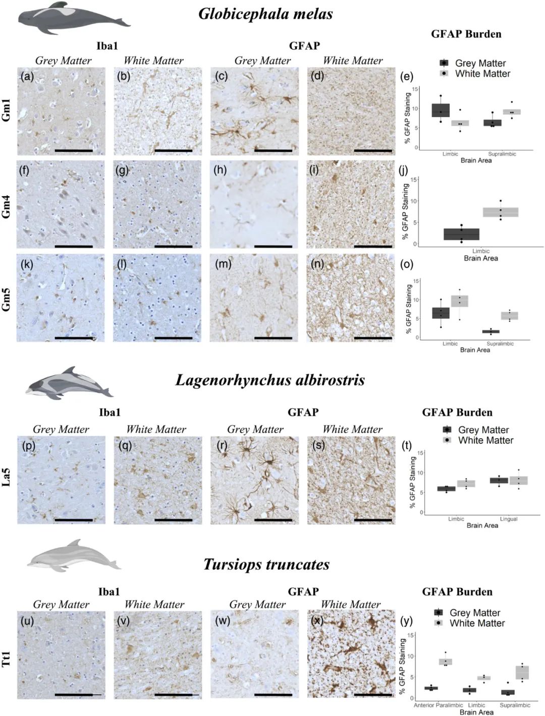
FIG5
Immunohistochemical labelling (brown pigment) of astrocytes and microglia in grey and white matter from Globicephala melas (a–o), Lagenorhynchus albirostris (p–t) and Tursiops truncatus (u–y). All animals have both Aβ and pTau pathology as assessed by IHC, with the exception of Gm4 whose pTau status was not determined. Due to sample availability, different brain regions were assessed for each animal, with GFAP burden values are reported as % area of the image occupied by GFAP labelling. Graphs show an average ±SEM of 5 200 × 200 μm randomly selected regions of interest from both grey and white matter in each brain region (e,j,o,t,y). Iba1 labelling was fairly weak in all animals, although individual cells were resolvable in some sections, notably white matter from La5 (q) and Tt1 (v). GFAP labelling was clearer, with individual cells clearly resolvable in grey matter from Gm1 (c), Gm4 (h), Gm5 (m) and La5 (r). Tt1 showed a different pattern, with highly diffuse labelling in grey matter (w), but more readily resolvable cells in white matter (x), in contrast to the more punctate/mesh-like GFAP labelling in white matter from other animals (d,i,n,s). Iba1 and GFAP labelling seemed uniformly distributed through grey/white matter, with little evidence of gliosis as may be seen around neuritic plaques in human Alzheimer's disease brain. Data are shown as box plots with each individual animal mean as data points. Scale bars = 100 μm
扫码阅读原文

https://onlinelibrary.wiley.com/doi/10.1111/ejn.15900
本网站所有内容来源注明为“梅斯医学”或“MedSci原创”的文字、图片和音视频资料,版权均属于梅斯医学所有。非经授权,任何媒体、网站或个人不得转载,授权转载时须注明来源为“梅斯医学”。其它来源的文章系转载文章,或“梅斯号”自媒体发布的文章,仅系出于传递更多信息之目的,本站仅负责审核内容合规,其内容不代表本站立场,本站不负责内容的准确性和版权。如果存在侵权、或不希望被转载的媒体或个人可与我们联系,我们将立即进行删除处理。
在此留言


学习了
0