


Cancers 2024, 16(9), 1671; https://doi.org/10.3390/cancers16091671 (registering DOI) - 25 Apr 2024
Abstract
Background: Chronic periodontitis, an inflammation-related disorder affecting global populations, has been revealed to be linked to diverse cancers. Numerous epidemiological studies have not shown a link between chronic periodontitis and blood cancers in Taiwan. Methods: This study included 601,628 patients, diagnosed with newly
[...] Read more.
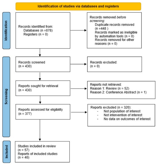
Background: Chronic periodontitis, an inflammation-related disorder affecting global populations, has been revealed to be linked to diverse cancers. Numerous epidemiological studies have not shown a link between chronic periodontitis and blood cancers in Taiwan. Methods: This study included 601,628 patients, diagnosed with newly chronic periodontitis by the ICD-9-CM classification, who were enrolled from 2001 to 2021 in the National Health Insurance Research Database (NHIRD) in Taiwan. In this study, we employed comprehensive statistical analyses to investigate the association between chronic periodontitis and hematologic cancers. Initially, we calculated incidence density and used a Poisson regression to analyze relative risk. Subsequently, we compared the cumulative incidence of hematological cancer in both chronic and non-chronic periodontitis groups using the Kaplan–Meier method. Results: The results revealed a significantly lower cumulative incidence of hematologic cancer in individuals with non-chronic periodontitis over a 12-year follow-up period. To further explore the risk factors, a Cox proportional hazard regression analysis was conducted. Being male (adjusted hazard ratio [aHR] = 1.21, 95% CI: 1.04 to 1.42; p = 0.014) and having hypertension (aHR = 1.34, 95% CI: 1.06 to 1.69; p = 0.015) were demonstrated to be associated with an increased risk of hematologic cancers, respectively. In addition, in a subtype multivariate analysis for categorizing hematologic cancers into lymphoma and leukemia, the aHR for leukemia was 1.48 (95% CI: 1.13 to 1.93; p = 0.004) and aHR for lymphoma was 1.15 (95% CI: 0.96 to 1.37; p = 0.140). Conclusions: This study found that being male and having hypertension were the significant risk factors for hematological malignancies. Moreover, the association between chronic periodontitis and specific subtypes of hematologic cancers was confirmed.
Full article
(This article belongs to the Special Issue Oral Potentially Malignant Disorders and Oral Cavity Cancer)


