


Int. J. Mol. Sci. 2024, 25(9), 4657; https://doi.org/10.3390/ijms25094657 (registering DOI) - 25 Apr 2024
Abstract
This study explores the impact of defecation frequency on the gut microbiome structure by analyzing fecal samples from individuals categorized by defecation frequency: infrequent (1–3 times/week, n = 4), mid-frequent (4–6 times/week, n = 7), and frequent (daily, n = 9). Utilizing 16S
[...] Read more.
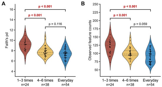
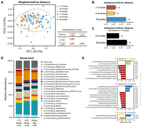
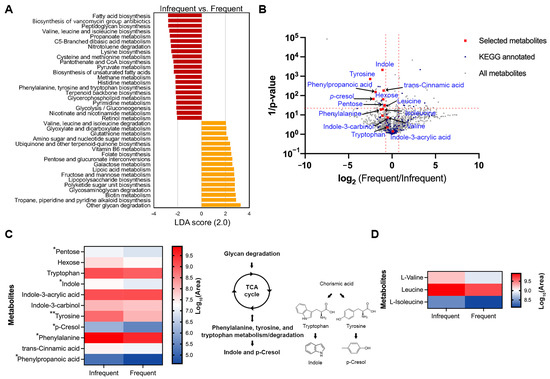
This study explores the impact of defecation frequency on the gut microbiome structure by analyzing fecal samples from individuals categorized by defecation frequency: infrequent (1–3 times/week, n = 4), mid-frequent (4–6 times/week, n = 7), and frequent (daily, n = 9). Utilizing 16S rRNA gene-based sequencing and LC-MS/MS metabolome profiling, significant differences in microbial diversity and community structures among the groups were observed. The infrequent group showed higher microbial diversity, with community structures significantly varying with defecation frequency, a pattern consistent across all sampling time points. The Ruminococcus genus was predominant in the infrequent group, but decreased with more frequent defecation, while the Bacteroides genus was more common in the frequent group, decreasing as defecation frequency lessened. The infrequent group demonstrated enriched biosynthesis genes for aromatic amino acids and branched-chain amino acids (BCAAs), in contrast to the frequent group, which had a higher prevalence of genes for BCAA catabolism. Metabolome analysis revealed higher levels of metabolites derived from aromatic amino acids and BCAA metabolism in the infrequent group, and lower levels of BCAA-derived metabolites in the frequent group, consistent with their predicted metagenomic functions. These findings underscore the importance of considering stool consistency/frequency in understanding the factors influencing the gut microbiome.
Full article
(This article belongs to the Special Issue New Molecular Insights into the Gut Microbiome)
►
Show Figures
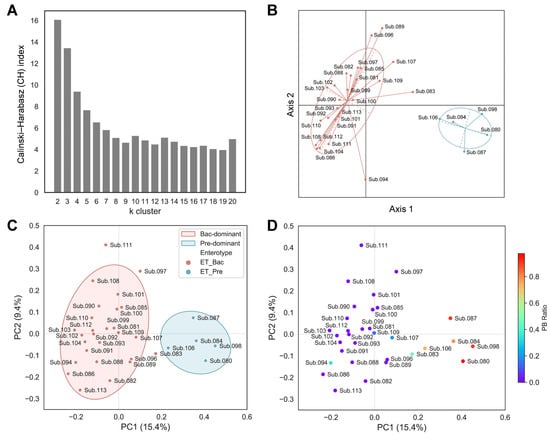
Figure 1


