【专家述评】| 2022年乳腺癌基础与转化研究进展
2023-08-03 中国癌症杂志 中国癌症杂志 发表于上海
本文从肿瘤代谢、肿瘤微环境、微生物与肿瘤、肿瘤转移、肿瘤耐药与药物筛选、多组学研究、人工智能共7个方面对2022年乳腺癌基础与转化研究的年度进展进行总结。
[摘要] 世界卫生组织(World Health Organization,WHO)国际癌症研究机构发布的数据显示,乳腺癌现已成为全球女性发病率最高的恶性肿瘤,严重威胁女性健康。2022年乳腺癌基础与转化研究领域进展颇丰,深化了对乳腺癌分子本质的认识,为乳腺癌精准治疗提供了新的思路。本文从肿瘤代谢、肿瘤微环境、微生物与肿瘤、肿瘤转移、肿瘤耐药与药物筛选、多组学研究、人工智能共7个方面对2022年乳腺癌基础与转化研究的年度进展进行总结,并对乳腺癌研究未来的发展方向进行展望,以期为乳腺癌研究的进一步深化提供参考。
[关键词] 乳腺肿瘤;基础与转化研究;研究进展
[Abstract] According to the data released by the International Cancer Research Institute of the World Health Organization (WHO), breast cancer is the malignant tumor with the highest incidence rate in women, and seriously endangers women's health. Many impressive advances have been achieved in fundamental and translational research of breast cancer, which deepened the understanding of the molecular nature of breast cancer and provided new scientific strategies for the precision treatment of breast cancer. This paper summarized the annual advances in fundamental and translational breast cancer research in 2022 in seven ps: tumor metabolism, tumor microenvironment, microbes and tumors, tumor metastasis, tumor drug resistance and drug screening, multi-omics research and artificial intelligence. Furthermore, we proposed an outlook on the future directions of breast cancer research to provide a reference for future studies.
[Key words] Breast cancer; Fundamental and translational research; Research advances
根据世界卫生组织国际癌症研究机构发布的数据显示,乳腺癌现已成为全球女性发病率最高的恶性肿瘤,严重威胁女性健康[1]。在过去的一年里,乳腺癌领域涌现了大量卓越的基础与转化研究成果,除了在既往研究较多的肿瘤代谢、肿瘤微环境、肿瘤转移和药物相关研究领域中不断推陈出新,近年来方兴未艾的微生物组学方面也有许多重要突破。此外,多组学研究的分析维度和研究广度不断扩大,人工智能也逐渐渗透至乳腺癌诊疗的各个环节。本文将总结归纳2022年乳腺癌基础与转化研究的最新进展,为乳腺癌的精准诊疗提供新的思路。
1 肿瘤代谢
2022年乳腺癌代谢领域的研究除了聚焦关键的能量代谢通路,也进一步向关键代谢物、铁死亡等领域拓展。在关键的葡萄糖代谢方面,Jiang等[2]证明Zeb1通过促进乳腺癌Warburg效应,诱导缺氧环境和乳酸产生,激活M2样巨噬细胞并导致肿瘤免疫抑制。而Zhu等[3]发现葡萄糖剥夺环境可以激活乳腺癌细胞的能量应激反应,导致能量代谢重编程和正反馈促癌信号。Chen等[4]则报道了长链非编码RNA(long non-coding RNA,lncRNA) DIO3OS通过稳定乳酸脱氢酶的信使RNA(message RNA,mRNA),上调乳腺癌糖酵解,并最终导致激素受体阳性乳腺癌对芳香化酶抑制剂的耐药性。
此外,多种代谢物的失调也被发现与肿瘤转移密切相关。脑转移乳腺癌细胞不同的乳酸和谷氨酰胺代谢特点决定了机体免疫活性和肿瘤细胞的转移能力[5]。乳腺癌细胞丙酸代谢失调导致甲基丙二酸的累积也能够增强乳腺癌的侵袭和转移能力[6]。Gong等[7]发现肺间充质细胞可以通过类脂转移的方式抑制自然杀伤(natural killer,NK)细胞功能并促进乳腺癌肺转移。
近年来,铁死亡作为一种与脂质过氧化物代谢功能障碍密切相关的非凋亡性细胞死亡方式[8-9],受到了众多学者的关注。在过去的一年里,在发现数个调控乳腺癌铁死亡的新型分子的同时[10-12],研究者也对乳腺癌铁死亡异质性和作用机制有了更深刻的理解。Yang等[13]通过大规模三阴性乳腺癌(triple-negative breast cancer,TNBC)多组学队列(n=465)剖析了TNBC的铁死亡异质性,并进一步发现腔面雄激素受体亚型(luminal androgen receptor,LAR)具有铁死亡相关代谢通路上调的特点,使用铁死亡诱导剂GPX4抑制剂可以有效地诱导LAR亚型的铁死亡并增敏免疫治疗。Zhang等[14]发现了乳腺癌细胞中铁死亡脂质过氧化代谢的信号转导放大通路,揭示了脂质过氧化-PKCβⅡ-ACSL4正反馈轴作为铁死亡相关肿瘤治疗靶点的潜在价值。Xie等[15]证实了乳腺脂肪细胞通过油酸保护乳腺癌细胞免于铁死亡的作用。此外,也有多项研究[16-19]通过设计纳米颗粒药物和工程红细胞,以扰乱脂质代谢,诱导铁死亡,从而起到治疗乳腺癌的作用。
2 肿瘤微环境
2.1 免疫细胞
肿瘤中浸润的多种免疫细胞构成了肿瘤微环境的重要组成部分,深入剖析肿瘤免疫细胞对于解构肿瘤免疫和肿瘤微环境有着深远的意义。在2022年,多种免疫细胞在乳腺癌中的作用得到了更加深入的挖掘。在抗肿瘤免疫中,CD4+T细胞通常被置于辅助地位,而Seung等[20]通过对靶向人表皮生长因子受体2(human epidermal growth factor receptor 2,HER2)、CD3、CD28的三特异性抗体的机制研究发现,CD4+T细胞可以直接阻断G1/S细胞周期,抑制HER2阳性乳腺癌细胞的生长,进一步拓展了人们对CD4+T细胞的认知。巨噬细胞同样是肿瘤免疫微环境的重要组成部分,但它在功能和表型上往往具有异质性。Nalio Ramos等[21]鉴定了一群FOLR2+组织驻留巨噬细胞,其通过与CD8+T细胞的密切接触激活抗肿瘤免疫,在激素受体阳性的乳腺癌中起到抑癌作用。而Nixon等[22]则发现肿瘤相关巨噬细胞增殖、成熟和抗原呈递所需的关键转录因子IRF8会促进细胞毒性T淋巴细胞(cytotoxic T lymphocyte,CTL)的耗竭,起到促癌作用。嗜酸性粒细胞在乳腺癌中的研究相对较少,但Blomberg等[23]发现免疫检查点抑制剂联合顺铂可以通过白介素(interleukin,IL)-5诱导嗜酸性粒细胞的扩增,并在IL-33的作用下促进嗜酸性粒细胞在乳腺肿瘤中的浸润,导致嗜酸性粒细胞依赖的CD8+T细胞活化,增强抗肿瘤免疫反应。
2.2 其他基质细胞与非细胞成分
除了免疫细胞外,肿瘤微环境中还存在肿瘤相关成纤维细胞(cancer-associated fibroblast,CAF)、内皮细胞等基质细胞及相关非细胞成分,它们也在肿瘤的增殖转移中发挥重要的调控作用。Kay等[24]发现CAF依赖于脯氨酸合成关键酶PYCR1产生促癌胶原蛋白,从而增强乳腺癌细胞的增殖和转移。CAF还可以通过分泌Ⅻ型胶原调控Ⅰ型胶原的累积,从而建立起促进转移的侵袭性肿瘤微环境[25]。而内皮细胞可以通过特定的分泌因子,形成促转移生态位,诱导乳腺癌细胞的干性表型和肺转移[26]。此外,乳腺脂肪细胞也可以去分化形成肌成纤维样细胞和巨噬细胞样细胞,从而起到促进肿瘤细胞增殖的作用[27]。因此,靶向肿瘤基质成为了一种颇具希望的乳腺癌治疗思路。Wang等[28]通过仿生脂蛋白药物递送系统靶向肿瘤基质,重塑肿瘤血管、降低CAF比例、清除胶原蛋白等细胞外基质成分,从而促进CTL的瘤内浸润。
在传统生物学作用之外,生物力学为我们提供了一种全新的解析肿瘤基质的视角[29]。Bera等[30]发现高细胞外黏度通过ARP-NHE1-TRPV4轴可重塑肿瘤细胞骨架,增强肿瘤细胞的迁移能力。而软的肿瘤基质也被证明可以通过肌动蛋白增加线粒体分裂,导致活性氧类增多,并进一步使得乳腺癌细胞免于化疗药物的杀伤[31]。
3 微生物与肿瘤
随着对肿瘤生态系统研究的深入,人们逐渐认识到多态微生物群作为一种肿瘤新特征的重要性[32]。既往研究[33-34]已经证实了细菌在乳腺肿瘤中的存在。在此基础上,2022年乳腺癌微生物研究聚焦于肿瘤内微生物和消化道微生物对乳腺癌发生、发展的影响,并向真菌组学发起了挑战。在肿瘤内微生物方面,Wang等[35]通过大规模多组学队列对TNBC中的微生物群组成分进行了分析,并发现免疫调节亚型(immunomodulatory)中富集的梭状芽胞杆菌代谢产物氧化三甲胺能够有效激活抗肿瘤免疫。Fu等[36]发现细胞内细菌能够重组循环肿瘤细胞的细胞骨架从而增强其存活能力,进而促进乳腺癌的转移。在消化道微生物方面,Deng等[37]发现肝螺杆菌可以从肠道向乳腺肿瘤移位,进而导致骨髓来源的抑制细胞在肿瘤组织的浸润,促进乳腺癌的生长。在真菌组学方面,两项大规模泛癌真菌组学研究[38-39]将微生物群组的研究维度进一步拓宽到真菌群组中,其发现马拉色菌属作为癌种特异性真菌在乳腺癌中显著富集,引起肿瘤内细菌-真菌-免疫相互作用,并能够有效区分患者的预后。研究者们也尝试利用微生物来治疗乳腺癌,Xiao等[40]利用厌氧菌对缺氧区域的亲和力,将厌氧菌作为阿霉素的运输载体,成功地将药物送往常规给药方法难以企及的缺氧区域,显著延长了乳腺癌小鼠的生存期。相信随着微生物在乳腺肿瘤中的作用被更深程度地认知,我们能够更好地利用微生物来治疗乳腺癌,与“菌”共舞。
4 肿瘤转移
肿瘤转移是导致乳腺癌患者死亡的主要原因之一,但目前转移的决定因素仍未探明。2022年,结合大型队列和单细胞测序等新数据、新技术,乳腺癌转移相关研究亮点频出。Nguyen等[41]通过大规模泛癌转移瘤队列(n=25 775)对肿瘤基因组特征与转移之间的相互作用进行了深入剖析,发掘出染色体不稳定性增高与乳腺癌转移负荷之间的密切关联。在表观遗传学方面,Garcia-Recio等[42]通过转移性乳腺癌队列发现,与原发灶相比,转移灶DNA甲基化介导细胞黏附基因表达下调,使浸润的免疫细胞数量减少,揭示了表观遗传学在乳腺癌转移中的重要调控作用。而肿瘤转移也离不开目标器官所形成的前转移生态位(pre-metastatic niche)。表达COX-2的肺成纤维细胞可以诱导肺髓系细胞的免疫抑制表型,形成前转移生态位[43]。同时,乳腺癌细胞也可以通过Lin28B上调N2型中性粒细胞,诱导肺前转移生态位的形成,从而促进乳腺癌细胞的肺转移[44]。
5 肿瘤耐药与药物筛选
5.1 耐药机制探索
肿瘤耐药是乳腺癌患者生存期进一步提升的主要障碍。2022年乳腺癌耐药相关研究从乳腺癌细胞和肿瘤微环境细胞两个角度出发揭示了耐药背后的深层机制。乳腺癌细胞可以通过形成“细胞内细胞”(cell-in-cell)和静止肿瘤细胞簇(quiescent cancer cells),或是转化为耐药持久性细胞(drug-tolerant persister cells,DTP)来逃逸药物的杀伤。Gutwillig等[45]发现,在干扰素(interferon,IFN)-γ作用下,乳腺癌细胞可以形成“细胞内细胞”结构,使得内层肿瘤细胞免受T细胞来源的穿孔素作用,从而产生免疫治疗抗性。Baldominos等[46]发现乳腺癌细胞可以形成静止肿瘤细胞簇,通过缺氧通路诱导T细胞功能耗竭、树突状细胞的功能失调和促癌CAF的浸润,最终导致免疫治疗耐药。Chang等[47]发现HER2阳性乳腺癌中具有前DTP,其在酪氨酸激酶抑制剂诱导下可以转化为DTP,并进一步通过AKT非依赖的mTORC1激活产生耐药性。此外,Marsolier等[48]发现阻止H3K27me3去甲基化可以防止肿瘤细胞进入DTP状态,显示表观遗传改变对乳腺癌细胞耐药性具有调控作用。
除了肿瘤细胞本身,微环境细胞也是介导肿瘤耐药的重要一环。Liu等[49]发现微环境中的CD16+ CAF可以通过FcγR与曲妥珠单抗相互作用,产生胶原沉积的微环境表型,影响药物运输,从而产生耐药性。患者体内的组胺可以诱导巨噬细胞向M2表型极化,并导致T细胞功能不良[50];缺氧也会诱导T细胞和NK细胞中效应基因的表观遗传学抑制[51],并最终导致乳腺癌免疫治疗耐药。
5.2 药物筛选
除了对已有耐药机制的深入探索,乳腺癌治疗药物筛选方面也有许多新进展。Jaaks等[52]通过在125种乳腺癌、结肠癌和胰腺癌细胞系中测量2 025种药物的两两组合效果,为探究药物的肿瘤治疗协同作用提供了丰富的数据支持。Guillen等[53]通过患者来源的异种移植物及其衍生的类器官模型(PDX-derived organoid)为临床患者筛选敏感药物提供了个性化治疗方案,实现了肿瘤精准治疗。
6 多组学研究
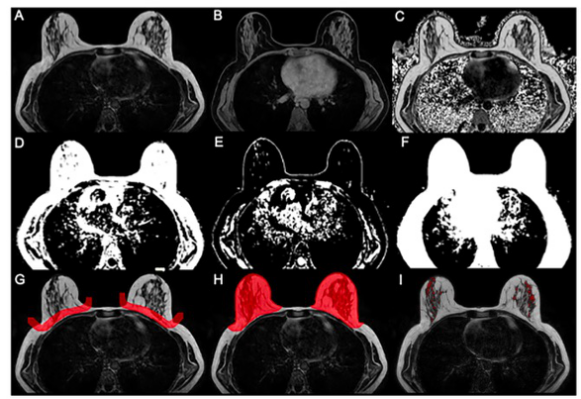
多组学研究已经成为一种全面解析肿瘤内在特征的重要手段,既往研究[54-59]通过基因组、转录组、蛋白质组等维度的数据对乳腺癌进行了全面分析。过去一年,乳腺癌领域的组学分析维度仍在不断扩展。在代谢组学方面,Xiao等[60]绘制了涵盖330例TNBC标本的大规模代谢组学图谱,将TNBC患者划分为3个代谢亚组,并通过配对多组学数据的整合分析,发现1-磷酸鞘氨醇和N-乙酰-天冬氨酸-谷氨酸分别为LAR亚型和高危基底样免疫抑制亚型(basal-like immune-suppressed subtype)的潜在靶点。而Jiang等[61]首次报道了涵盖影像组学的大规模TNBC临床队列,通过影像组学和其他组学维度的整合分析,深入探索了影像组学特征与TNBC肿瘤异质性、代谢重编程、肿瘤微环境之间的关联,并成功通过影像组学数据对TNBC分子亚型进行预测。除此之外,空间基因组学[62]和单细胞基因组测序技术[63]也被用于从单细胞及空间维度揭示乳腺癌的克隆进化和基因组突变模式,进一步丰富了乳腺癌多组学研究的维度。
在分析维度不断扩展的同时,多组学研究进一步触及更多人种和更多乳腺肿瘤亚型的分子本质。Martini等[64]基于大规模非洲裔美国人及非洲TNBC队列,通过全面的遗传血统评估发现了非洲裔血统相关基因,从而揭示了非洲裔血统与TNBC免疫异质性之间的关联。而作为侵袭性乳腺癌的前兆,导管原位癌(ductal carcinoma in situ,DCIS)的分子本质和微环境特征也在过去一年中被深入剖析,研究者通过多组学及单细胞数据[65-66]和基于MIBI-TOF的空间细胞图谱[67],明确了高风险DCIS的基因和微环境特征,分析了DCIS与复发侵袭性乳腺癌之间的克隆关系,加深了我们对DCIS的理解。
7 人工智能
伴随着算法的迭代,人工智能已深入到乳腺癌诊疗的全程,并在过去的一年里获得了众多突破性进展。在乳腺癌的早期筛查方面,Yala等[68]通过人工智能驱动的风险模型Tempo,在提高乳腺癌早期筛查能力的同时降低了筛查成本。在乳腺癌诊断分级方面,Wang等[69]基于数字病理学数据通过深度学习算法改进了乳腺癌组织学分级,可有效地区分高危患者。在预测治疗反应方面,Sammut等[70]通过机器学习算法整合了多组学特征,可有效地预测乳腺癌患者的治疗反应(AUC=0.87)。在综合运用方面,Zhao等[71]使用多组学TNBC队列(n=425),设计了深度学习算法框架,使用数字病理数据综合预测了TNBC患者的分子特征、分子亚型和临床预后。可以预见在不远的将来,人工智能将真正走进临床,并在乳腺癌早期筛查、诊断分级、分子分型和疗效预测方面发挥巨大的作用。
8 总结与展望
2022年乳腺癌基础与转化研究领域取得了重要进展,但仍面临许多挑战。多项研究揭示了铁死亡在乳腺癌中的异质性和作用机制,使人们对铁死亡有了更加清晰的认识。乳腺癌肿瘤微环境研究更加精细化,利用单细胞测序工具,揭示了不同微观亚群乳腺癌的生物学特征。乳腺癌微生物研究已然扣响了真菌组学的大门,但不同真菌种属在乳腺癌中的具体作用及其机制仍有待进一步探索。人工智能算法已经展示出潜在的临床运用价值,但仍需要前瞻性临床队列验证其解决复杂临床问题的能力。期待在不久的将来,更多的基础与转化研究能够为我们提供这些问题的答案并为临床决策提供参考,为乳腺癌患者带来更多获益。
利益冲突声明:所有作者均声明不存在利益冲突。
[参考文献]
[1] SUNG H, FERLAY J, SIEGEL R L, et al. Global cancer statistics 2020: GLOBOCAN estimates of incidence and mortality worldwide for 36 cancers in 185 countries[J]. CA Cancer J Clin, 2021, 71(3): 209-249.
[2] JIANG H M, WEI H M, WANG H, et al. Zeb1-induced metabolic reprogramming of glycolysis is essential for macrophage polarization in breast cancer[J]. Cell Death Dis, 2022, 13(3): 206.
[3] ZHU W J, C HE N X, GU O X Y, e t a l . Lo w g l u c o se - induced overexpression of HOXC-AS3 promotes metabolic reprogramming of breast cancer[J]. Cancer Res, 2022, 82(5): 805-818.
[4] CHEN X M, LUO R, ZHANG Y M, et al. Long noncoding RNA DIO3OS induces glycolytic-dominant metabolic reprogramming to promote aromatase inhibitor resistance in breast cancer[J]. Nat Commun, 2022, 13(1): 7160.
[5] PARIDA P K, MARQUEZ-PALENCIA M, NAIR V, et al. Metabolic diversity within breast cancer brain-tropic cells determines metastatic fitness[J]. Cell Metab, 2022, 34(1): 90-105.e7.
[6] GOMES A P, ILTER D, LOW V, et al. Altered propionate metabolism contributes to tumour progression and aggressiveness[J]. Nat Metab, 2022, 4(4): 435-443.
[7] GONG Z, LI Q, SHI J Y, et al. Lipid-laden lung mesenchymal cells foster breast cancer metastasis via metabolic reprogramming of tumor cells and natural killer cells[J]. Cell Metab, 2022, 34(12): 1960-1976.e9.
[8] DIXON S J, LEMBERG K M, LAMPRECHT M R, et al. Ferroptosis: an iron-dependent form of nonapoptotic cell death[J]. Cell, 2012, 149(5): 1060-1072.
[9] TANG D L, CHEN X, KANG R, et al. Ferroptosis: molecular mechanisms and health implications[J]. Cell Res, 2021, 31(2): 107-125.
[10] LI H Y, YANG P H, WANG J H, et al. HLF regulates ferroptosis, development and chemoresistance of triple-negative breast cancer by activating tumor cell-macrophage crosstalk[J]. J Hematol Oncol, 2022, 15(1): 2.
[11] WU M M, ZHANG X, ZHANG W J, et al. Cancer stem cell regulated phenotypic plasticity protects metastasized cancer cells from ferroptosis[J]. Nat Commun, 2022, 13(1): 1371.
[12] ZOU Y T, ZHENG S Q, XIE X H, et al. N6-methyladenosine regulated FGFR4 attenuates ferroptotic cell death in recalcitrant HER2-positive breast cancer[J]. Nat Commun, 2022, 13(1): 2672.
[13] YANG F, XIAO Y, DING J H, et al. Ferroptosis heterogeneity in triple-negative breast cancer reveals an innovative immunotherapy combination strategy[J]. Cell Metab, 2023, 35(1): 84-100.e8.
[14] ZHANG H L, HU B X, LI Z L, et al. PKCβⅡ phosphorylates ACSL4 to amplify lipid peroxidation to induce ferroptosis[J]. Nat Cell Biol, 2022, 24(1): 88-98.
[15] XIE Y Z, WANG B Y, ZHAO Y N, et al. Mammary adipocytes protect triple-negative breast cancer cells from ferroptosis[J]. J Hematol Oncol, 2022, 15(1): 72.
[16] LI K, LIN C C, LI M H, et al. Multienzyme-like reactivity cooperatively impairs glutathione peroxidase 4 and ferroptosis suppressor protein 1 pathways in triple-negative breast cancer for sensitized ferroptosis therapy[J]. ACS Nano, 2022, 16(2): 2381-2398.
[17] YANG J, JIA Z G, ZHANG J, et al. Metabolic intervention nanoparticles for triple-negative breast cancer therapy via overcoming FSP1-mediated ferroptosis resistance[J]. Adv Healthc Mater, 2022, 11(13): e2102799.
[18] PAN W L, TAN Y, MENG W, et al. Microenvironment-driven sequential ferroptosis, photodynamic therapy, and chemotherapy for targeted breast cancer therapy by a cancer-cell-membranecoated nanoscale metal-organic framework[J]. Biomaterials, 2022, 283: 121449.
[19] ZHOU A W, FANG T L, CHEN K R, et al. Biomimetic activator of sonodynamic ferroptosis amplifies inherent peroxidation for improving the treatment of breast cancer[J]. Small, 2022, 18(12): e2106568.
[20] SEUNG E, XING Z, WU L, et al. A trispecific antibody targeting HER2 and T cells inhibits breast cancer growth via CD4 cells[J]. Nature, 2022, 603(7900): 328-334.
[21] NALIO RAMOS R, MISSOLO-KOUSSOU Y, GERBERFERDER Y, et al. Tissue-resident FOLR2+ macrophages associate with CD8+ T cell infiltration in human breast cancer[J]. Cell, 2022, 185(7): 1189-1207.e25.
[22] NIXON B G, KUO F S, JI L L, et al. Tumor-associated macrophages expressing the transcription factor IRF8 promote T cell exhaustion in cancer[J]. Immunity, 2022, 55(11): 2044- 2058.e5.
[23] BLOMBERG O S, SPAGNUOLO L, GARNER H, et al. IL-5- producing CD4+ T cells and eosinophils cooperate to enhance response to immune checkpoint blockade in breast cancer[J]. Cancer Cell, 2023, 41(1): 106-123.e10.
[24] KAY E J, PATERSON K, RIERA-DOMINGO C, et al. Cancerassociated fibroblasts require proline synthesis by PYCR1 for the deposition of pro-tumorigenic extracellular matrix[J]. Nat Metab, 2022, 4(6): 693-710.
[25] PAPANICOLAOU M, PARKER A L, YAM M, et al. Temporal profiling of the breast tumour microenvironment reveals collagen Ⅻ as a driver of metastasis[J]. Nat Commun, 2022, 13(1): 4587.
[26] HONGU T, PEIN M, INSUA-RODRÍGUEZ J, et al. Perivascular tenascin C triggers sequential activation of macrophages and endothelial cells to generate a pro-metastatic vascular niche in the lungs[J]. Nat Cancer, 2022, 3(4): 486-504.
[27] ZHU Q Z, ZHU Y, HEPLER C, et al. Adipocyte mesenchymal transition contributes to mammary tumor progression[J]. Cell Rep, 2022, 40(11): 111362.
[28] WANG Y Q, XIE H L, WU Y, et al. Bioinspired lipoproteins of furoxans-oxaliplatin remodel physical barriers in tumor to potentiate T-cell infiltration[J]. Adv Mater, 2022, 34(14): e2110614.
[29] NIA H T, MUNN L L, JAIN R K. Physical traits of cancer[J]. Science, 2020, 370(6516): eaaz0868.
[30] BERA K, KIEPAS A, GODET I, et al. Extracellular fluid viscosity enhances cell migration and cancer dissemination[J]. Nature, 2022, 611(7935): 365-373.
[31] ROMANI P, NIRCHIO N, ARBOIT M, et al. Mitochondrial fission links ECM mechanotransduction to metabolic redox homeostasis and metastatic chemotherapy resistance[J]. Nat Cell Biol, 2022, 24(2): 168-180.
[32] HANAHAN D. Hallmarks of cancer: new dimensions[J]. Cancer Discov, 2022, 12(1): 31-46.
[33] HIEKEN T J, CHEN J, HOSKIN T L, et al. The microbiome of aseptically collected human breast tissue in benign and malignant disease[J]. Sci Rep, 2016, 6: 30751.
[34] NEJMAN D, LIVYATAN I, FUKS G, et al. The human tumor microbiome is composed of tumor type-specific intracellular bacteria[J]. Science, 2020, 368(6494): 973-980.
[35] WANG H, RONG X Y, ZHAO G, et al. The microbial metabolite trimethylamine N-oxide promotes antitumor immunity in triplenegative breast cancer[J]. Cell Metab, 2022, 34(4): 581-594. e8.
[36] FU A K, YAO B Q, DONG T T, et al. Tumor-resident intracellular microbiota promotes metastatic colonization in breast cancer[J]. Cell, 2022, 185(8): 1356-1372.e26.
[37] DENG H, MUTHUPALANI S, ERDMAN S, et al. Translocation of helicobacter hepaticus synergizes with myeloid-derived suppressor cells and contributes to breast carcinogenesis[J]. Oncoimmunology, 2022, 11(1): 2057399.
[38] NARUNSKY-HAZIZA L, SEPICH-POORE G D, LIVYATAN I, et al. Pan-cancer analyses reveal cancer-type-specific fungal ecologies and bacteriome interactions[J]. Cell, 2022, 185(20): 3789-3806.e17.
[39] DOHLMAN A B, KLUG J, MESKO M, et al. A pancancer mycobiome analysis reveals fungal involvement in gastrointestinal and lung tumors[J]. Cell, 2022, 185(20): 3807-3822.e12.
[40] XIAO S S, SHI H, ZHANG Y, et al. Bacteria-driven hypoxia targeting delivery of chemotherapeutic drug proving outcome of breast cancer[J]. J Nanobiotechnol, 2022, 20(1): 178.
[41] N G U Y E N B , F O N G C , L U T H R A A , e t a l . G e n o m i c characterization of metastatic patterns from prospective clinical sequencing of 25, 000 patients[J]. Cell, 2022, 185(3): 563-575.e11.
[42] GARCIA-RECIO S, HINOUE T, WHEELER G L, et al. Multiomics in primary and metastatic breast tumors from the AURORA US network finds microenvironment and epigenetic drivers of metastasis[J]. Nat Cancer, 2022[Epub ahead of print].
[43] GONG Z, LI Q, SHI J Y, et al. Lung fibroblasts facilitate premetastatic niche formation by remodeling the local immune microenvironment[J]. Immunity, 2022, 55(8): 1483-1500.e9.
[44] QI M Y, XIA Y, WU Y J, et al. Lin28B-high breast cancer cells promote immune suppression in the lung pre-metastatic niche via exosomes and support cancer progression[J]. Nat Commun, 2022, 13(1): 897.
[45] GUTWILLIG A, SANTANA-MAGAL N, FARHAT-YOUNIS L, et al. Transient cell-in-cell formation underlies tumor relapse and resistance to immunotherapy[J]. Elife, 2022, 11: e80315.
[46] BALDOMINOS P, BARBERA-MOURELLE A, BARREIRO O, et al. Quiescent cancer cells resist T cell attack by forming an immunosuppressive niche[J]. Cell, 2022, 185(10): 1694- 1708.e19.
[47] CHANG C A, JEN J, JIANG S W, et al. Ontogeny and vulnerabilities of drug-tolerant persisters in HER2+ breast cancer[J]. Cancer Discov, 2022, 12(4): 1022-1045.
[48] MARSOLIER J, PROMPSY P, DURAND A, et al. H3K27me3 conditions chemotolerance in triple-negative breast cancer[J]. Nat Genet, 2022, 54(4): 459-468.
[49] LIU X W, LU Y W, HUANG J Y, et al. CD16+ fibroblasts foster a trastuzumab-refractory microenvironment that is reversed by VAV2 inhibition[J]. Cancer Cell, 2022, 40(11): 1341-1357. e13.
[50] LI H Z, XIAO Y, LI Q, et al. The allergy mediator histamine confers resistance to immunotherapy in cancer patients via activation of the macrophage histamine receptor H1[J]. Cancer Cell, 2022, 40(1): 36-52.e9.
[51] MA S J, ZHAO Y, LEE W C, et al. Hypoxia induces HIF1α-dependent epigenetic vulnerability in triple-negative breast cancer to confer immune effector dysfunction and resistance to anti-PD-1 immunotherapy[J]. Nat Commun, 2022, 13(1): 4118.
[52] JAAKS P, COKER E A, VIS D J, et al. Effective drug combinations in breast, colon and pancreatic cancer cells[J]. Nature, 2022, 603(7899): 166-173.
[53] GUILLEN K P, FUJITA M, BUTTERFIELD A J, et al. A human breast cancer-derived xenograft and organoid platform for drug discovery and precision oncology[J]. Nat Cancer, 2022, 3(2): 232-250.
[54] JIANG Y Z, MA D, SUO C, et al. Genomic and transcriptomic landscape of triple-negative breast cancers: subtypes and treatment strategies[J]. Cancer Cell, 2019, 35(3): 428-440. e5.
[55] BERTUCCI F, NG C K Y, PATSOURIS A, et al. Genomic characterization of metastatic breast cancers[J]. Nature, 2019, 569(7757): 560-564.
[56] MERTINS P, MANI D R, RUGGLES K V, et al. Proteogenomics connects somatic mutations to signalling in breast cancer[J]. Nature, 2016, 534(7605): 55-62.
[57] CURTIS C, SHAH S P, CHIN S F, et al. The genomic and transcriptomic architecture of 2 000 breast tumours reveals novel subgroups[J]. Nature, 2012, 486(7403): 346-352.
[58] PEREIRA B, CHIN S F, RUEDA O M, et al. Erratum: the somatic mutation profiles of 2, 433 breast cancers refine their genomic and transcriptomic landscapes[J]. Nat Commun, 2016, 7: 11908.
[59] BARECHE Y, VENET D, IGNATIADIS M, et al. Unravelling triple-negative breast cancer molecular heterogeneity using an integrative multiomic analysis[J]. Ann Oncol, 2018, 29(4): 895-902.
[60] XIAO Y, MA D, YANG Y S, et al. Comprehensive metabolomics expands precision medicine for triple-negative breast cancer[J]. Cell Res, 2022, 32(5): 477-490.
[61] JIANG L, YOU C, XIAO Y, et al. Radiogenomic analysis reveals tumor heterogeneity of triple-negative breast cancer[J]. Cell Rep Med, 2022, 3(7): 100694.
[62] LOMAKIN A, SVEDLUND J, STRELL C, et al. Spatial genomics maps the structure, nature and evolution of cancer clones[J]. Nature, 2022, 611(7936): 594-602.
[63] FUNNELL T, O'FLANAGAN C H, WILLIAMS M J, et al. Single-cell genomic variation induced by mutational processes in cancer[J]. Nature, 2022, 612(7938): 106-115.
[64] MARTINI R, DELPE P, CHU T R, et al. African ancestryassociated gene expression profiles in triple-negative breast cancer underlie altered tumor biology and clinical outcome in women of African descent[J]. Cancer Discov, 2022, 12(11): 2530-2551.
[65] STRAND S H, RIVERO-GUTIÉRREZ B, HOULAHAN K E, et al. Molecular classification and biomarkers of clinical outcome in breast ductal carcinoma in situ: analysis of TBCRC 038 and RAHBT cohorts[J]. Cancer Cell, 2022, 40(12): 1521-1536. e7.
[66] LIPS E H, KUMAR T, MEGALIOS A, et al. Genomic analysis defines clonal relationships of ductal carcinoma in situ and recurrent invasive breast cancer[J]. Nat Genet, 2022, 54(6): 850-860.
[67] RISOM T, GLASS D R, AVERBUKH I, et al. Transition to invasive breast cancer is associated with progressive changes in the structure and composition of tumor stroma[J]. Cell, 2022, 185(2): 299-310.e18.
[68] YALA A, MIKHAEL P G, LEHMAN C, et al. Optimizing riskbased breast cancer screening policies with reinforcement learning[J]. Nat Med, 2022, 28(1): 136-143.
[69] WANG Y, ACS B, ROBERTSON S, et al. Improved breast cancer histological grading using deep learning[J]. Ann Oncol, 2022, 33(1): 89-98.
[70] SAMMUT S J, CRISPIN-ORTUZAR M, CHIN S F, et al. Multiomic machine learning predictor of breast cancer therapy response[J]. Nature, 2022, 601(7894): 623-629.
[71] ZHAO S, YAN C Y, LV H, et al. Deep learning framework for comprehensive molecular and prognostic stratifications of triple-negative breast cancer[J]. Fundam Res, 2022[Epub ahead of print].
本网站所有内容来源注明为“梅斯医学”或“MedSci原创”的文字、图片和音视频资料,版权均属于梅斯医学所有。非经授权,任何媒体、网站或个人不得转载,授权转载时须注明来源为“梅斯医学”。其它来源的文章系转载文章,或“梅斯号”自媒体发布的文章,仅系出于传递更多信息之目的,本站仅负责审核内容合规,其内容不代表本站立场,本站不负责内容的准确性和版权。如果存在侵权、或不希望被转载的媒体或个人可与我们联系,我们将立即进行删除处理。
在此留言


