

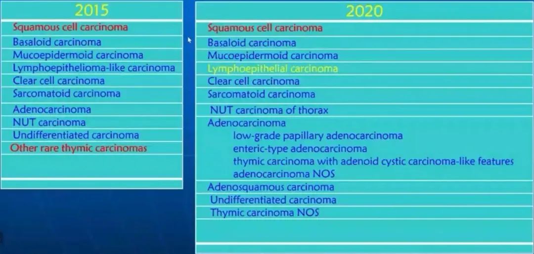
内容摘要
一、胸腺瘤(thymoma)
WHO“胸腺瘤病理分类”:
Type A thymoma:
TdT(+) T-cells in 10% or less of the tumor area are considered as type A thymomas
atypical type A thymoma
-
“atypia” with mitotic activity (≧ 4/10HPF)
-
“true” (coagulation) tumor necrosistrue
Controversial criteria:
Hypercellularity
enlarged hyperchromatic nuclei
large nucleoli, small mucous cysts
increased Ki67 index
extent of atypical areas
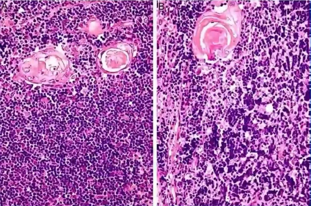
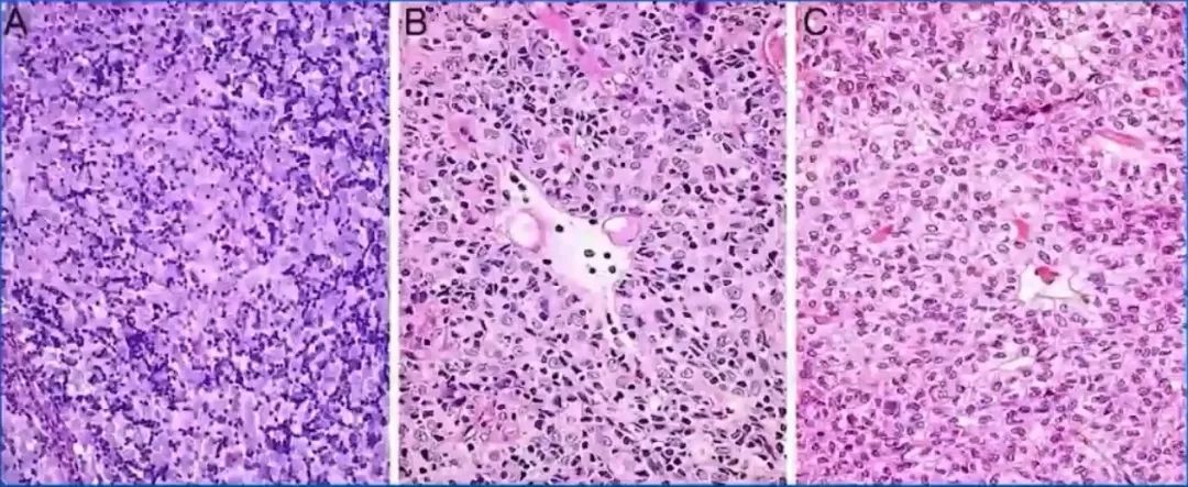
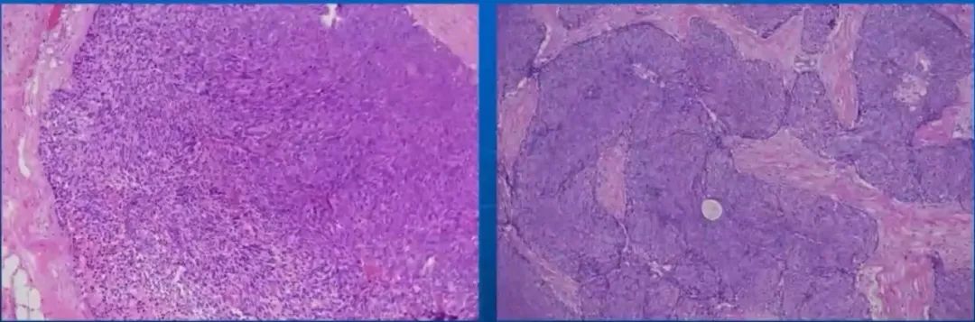
Atypical type A thymoma
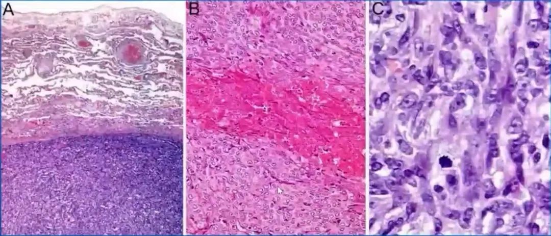
A.Pulmonary metastasis of type A thymoma (H&E, x50);
B.Spindle cell type A with comedo-type necrosis (H&E, x200);
C.Mitotic activity in a more epithelioid area of type A thymoma (H&E, x400)
Type AB thymoma:

Type B1 thymoma:
Type B2 thymoma:
Type B3 thymoma:
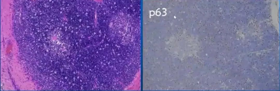
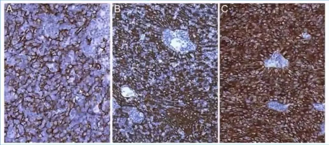
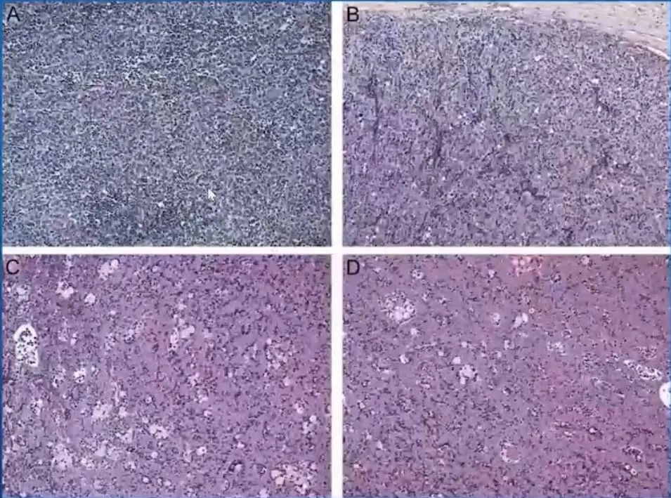
Type B1 thymoma
A.Organoid architecture with medullary island and preponderance ofdarker staining cortical area;
B.TDT expression by immature T-cells almost exclusively inthe cortical area;
C.Cytokeratin expression (antibody AE1/3) by a loose network ofepithelial cells in the cortical and medullary areas; by contrast, perivascular spaces aredistinctly epithelial free ;
D.Low number of evenly spaced neoplastic thymic epithelial cells as highlighted by p63 staining of tumor cell nuclei.
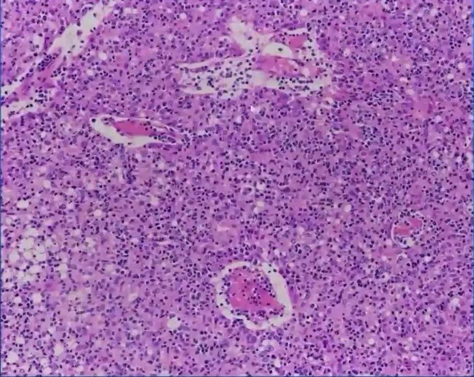
Hassall's corpuscles as optional feature in type B1 and B2 thvmoma
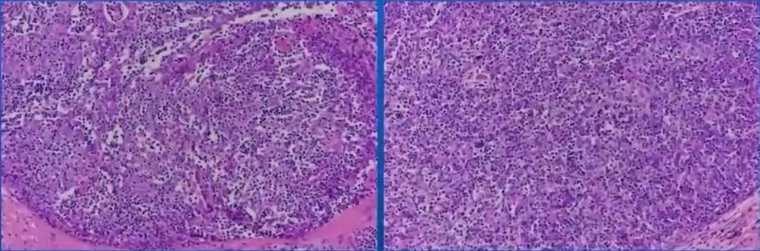
A.lymphocyte-rich type B1 thymoma with almost no discernible epithelial cells;
B.more epithelial-rich type B2 thymoma with easily discernable neoplastic epithelialcells.
Density of epithelial cell networks and delineation of perivascular spaces(PVS) as helpful features to distinguish B1, B2 and B3 thymomas.
A.Loose epithelial cell network and inconspicuous PVS in B1 thymoma;
B.Distinctly denser epithelial cell network and conspicuous PVS in type B2 thymoma;
C.Even stronger epithelial cell staining in B3 thymoma (pan-cytokeratin antibody AE1/3, X200).
B1 versus B2 thymoma, spectrum of "cortical areas”
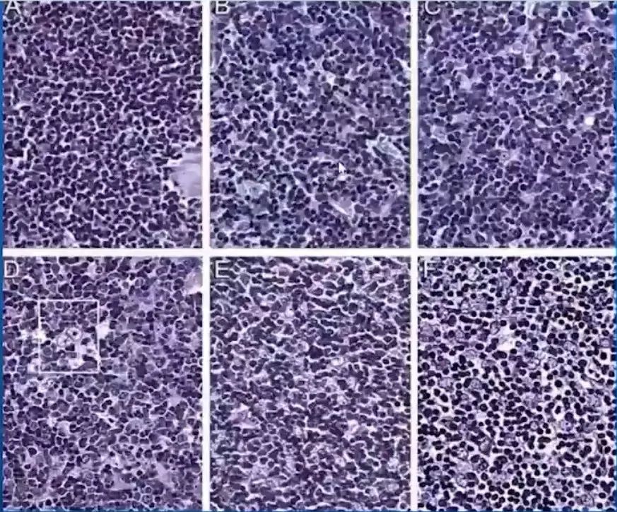
classical (A-B) and more epithelial-rich (C-E) B1 thymomas, "borderland" B1/B2 cases with too dense (F) or incipient clustering (G) of epithelial cells.
classical (H-L) 82 ttrymamas with increasing numbers of clustered epithelial cells.
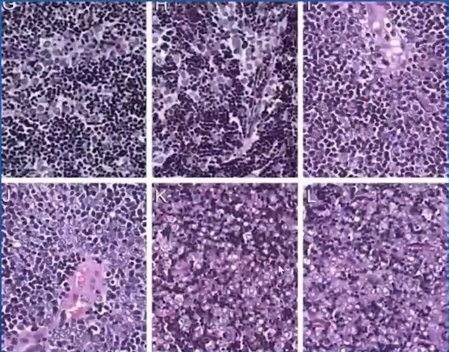
Distinction between type B2 and B3 thymomas
A.B2 thymomas:typically impression of a "blue "staining tumor on H&E histology due to the high content of lymphocytes;
B. and C.B3 thymoma: impression of a"pink"staining tumor due to the (variable) paucity of lymphocytes and abundance of slightly eosinophilic or clear epithelila cells (H&E, X200).
B2 versus B3 thymoma
classical (A-B) B2 thymomas,borderland B2/B3 cases (C-D).
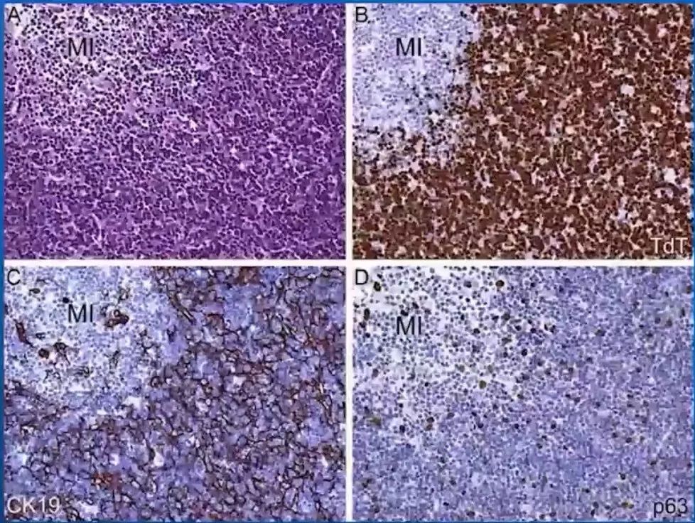
P63
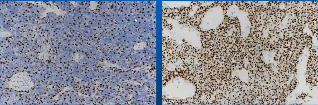
左图B2,右图B3
Micronodular thymoma:
Metaplastic thymoma:

二、胸腺癌(thymic carcinoma)
WHO“胸腺癌病理分类”:
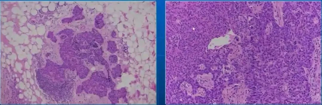
Squamous cell Carcinoma,SQ:
“B3/TC borderland tumors”
looked like B3 thymomas on H&E but showed two features of TC,namely CD5/CD117 expression and lack of TdT+T cells.
Keratinized SQ;+B3:
Spindle cell SQ:
Sarcomatoid carcinoma:

Mucoepidermoid carcinoma:

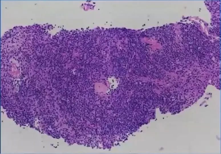
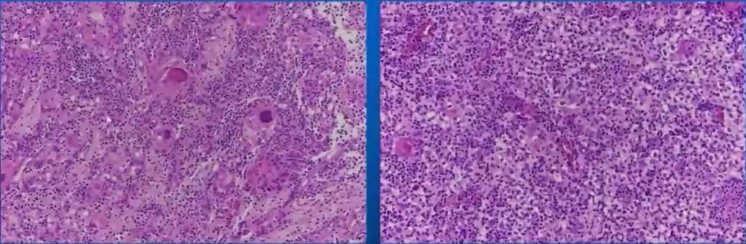
Lymphoepithelial carcinoma:
PD-L1 and C-KIT (could represent targets of potential therapeutic use.)
CLIC2 and SMAD4 as potential diagnostic biomarkers of thymic non-keratinized squamous cell carcinoma screened by iTRAQ.
诊断要点
A型胸腺瘤(同义词:梭形细胞),无器官样结构
但:含梭形细胞的TET≠A型胸腺瘤
鉴别诊断·梭形细胞变异型B3、类癌、梭形细胞性鳞癌;胸腺瘤化疗后改变。
AB型胸腺瘤
B型区域与B1/B2/B3的概念不同。
B1型胸腺瘤
瘤细胞散在分布,不成串。
B2型胸腺瘤
瘤细胞成串,与淋巴细胞各占一半。常伴有MG;肿瘤细胞围绕血管周隙成栅栏状排列。
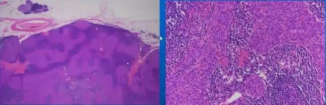
B3型胸腺瘤与鳞癌
B3型肿瘤细胞呈鳞片状排列;鳞癌细胞呈巢状,内部无未成熟T淋巴细胞浸润。
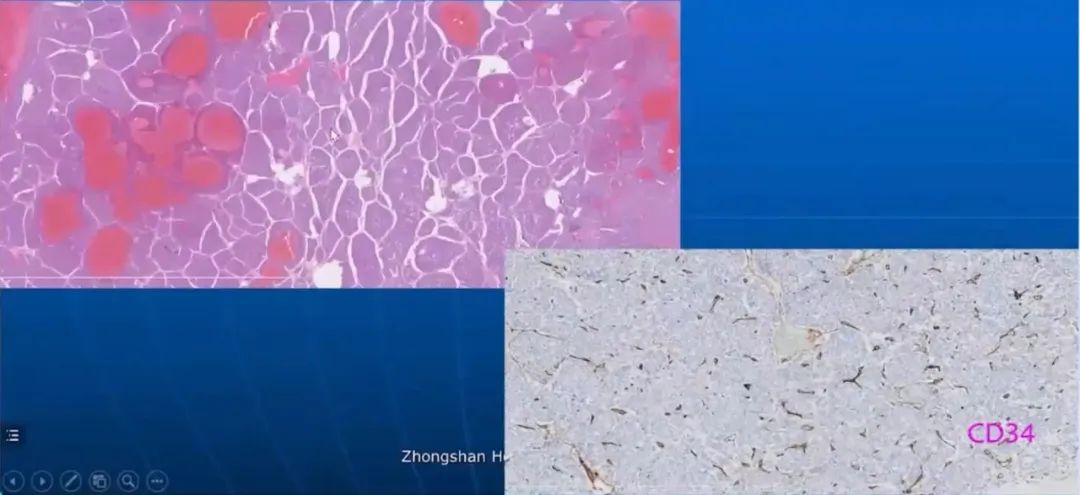
三、胸腺神经内分泌肿瘤(thymic neuroendocrine neoplasms,TNENs)
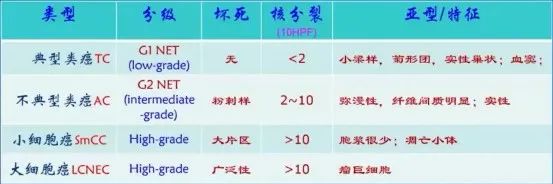
typical carcinoid:
atypical carcinoid:
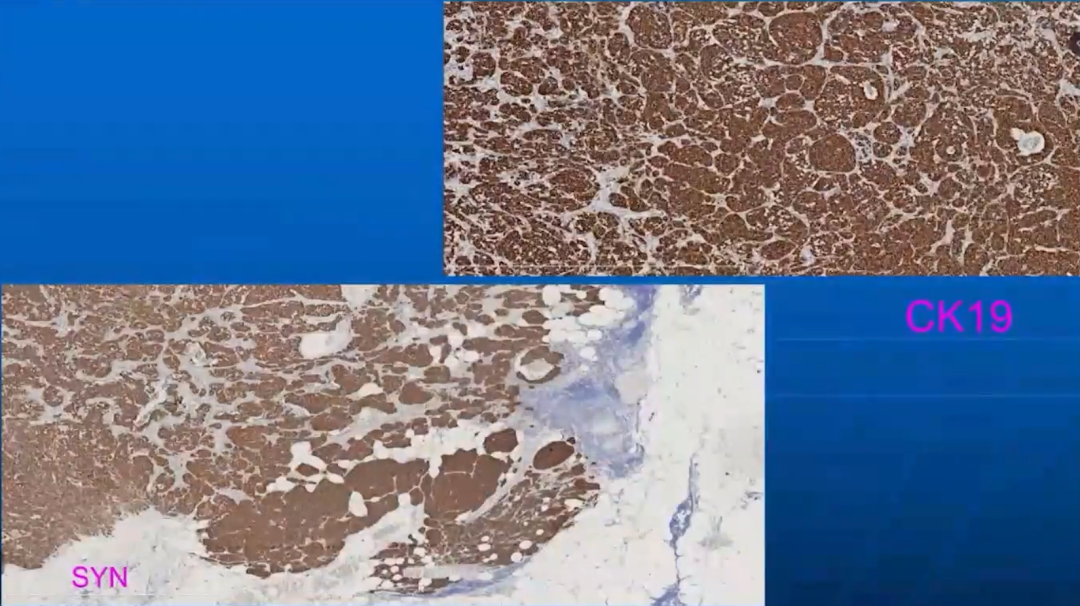
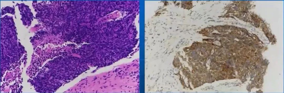
Large cell neuroendocrine carcinoma:
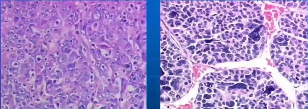
Small cell carcinoma:
本网站所有内容来源注明为“梅斯医学”或“MedSci原创”的文字、图片和音视频资料,版权均属于梅斯医学所有。非经授权,任何媒体、网站或个人不得转载,授权转载时须注明来源为“梅斯医学”。其它来源的文章系转载文章,或“梅斯号”自媒体发布的文章,仅系出于传递更多信息之目的,本站仅负责审核内容合规,其内容不代表本站立场,本站不负责内容的准确性和版权。如果存在侵权、或不希望被转载的媒体或个人可与我们联系,我们将立即进行删除处理。
在此留言