胃神经内分泌肿瘤的诊治现状与争议

2023-09-21 崔婷, 李金洲, 雷霞, 李继昂, 冯洁, 黄晓俊 协和医学杂志 发表于上海

神经内分泌肿瘤(neuroendocrine neoplasms, NENs)是起源于神经内分泌细胞和肽能神经元的高度异质性肿瘤,发病率正逐年提升

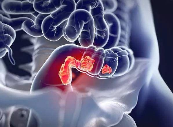
神经内分泌肿瘤(neuroendocrine neoplasms, NENs)是起源于神经内分泌细胞和肽能神经元的高度异质性肿瘤,发病率正逐年提升[1]。据美国监测、流行病学和最终结果(Surveillance, Epidemiology, and End Results, SEER)数据库统计显示,其发病率从2004年的5.25/10万上升至2012年的6.98/10万[2]。胃神经内分泌肿瘤(gastric neuroendocrine neoplasms, G-NENs)约占所有NENs的7.0%,在胃肿瘤中占比不足1.0%[3]。G-NENs发病率低且存在高度异质性,不同类型具有不同的诊治方式及预后。近年来,随着对该病认识的提高,国内外相关临床研究逐渐增多,且在临床诊断及治疗方面取得了巨大进展。值得注意的是,目前该病诊治仍存在较多争议,因此,本文就G-NENs在临床诊治中的最新研究进展进行综述,并对现存争议进行讨论。

近年来,有关G-NENs的临床分型一直存在争议,欧洲神经内分泌协会(European Neuroendocrine Tumor Society, ENETS)、北美神经内分泌肿瘤学会(North American Neuroendocrine Tumor Society, NANETS) 以及美国国家综合癌症网络(National Comprehensive Cancer Network, NCCN) 均建议将G-NENs分为三型。其中,Ⅰ型最为常见,约占所有病变的70.0%~80.0%,与慢性自身免疫性萎缩性胃炎引起的胃泌素分泌过量、肠嗜铬细胞增生/肥大有关;Ⅱ型约占所有病变的5.0%~10.0%,与胃泌素瘤或多发性内分泌腺瘤病1型(multiple endocrine neoplasia type 1,MEN1) 分泌的大量胃泌素导致的肠嗜铬细胞增生有关;Ⅲ型为散发性肿瘤,与任何已知疾病无关,发病机制尚未明确,约占所有病变的10.0%~15.0%[4-5]。有关Ⅲ型G-NENs的定义一直是争议的焦点。2006版ENETS指南将Ⅲ型G-NENs定义为分化良好/中等、非Ⅰ、Ⅱ型且无背景疾病的G-NENs,其定义与2010版、2013版NANETS指南以及2016版NCCN指南一致[6]。然而,2012版ENETS指南指出,Ⅲ型G-NENs的组织学类型多为分化较差的神经内分泌癌(neuroendocrine carcinoma, NEC)[7],基于此,对于分化良好的非Ⅰ型和非Ⅱ型的G-NENs将无法进行临床分类,而在2016版ENETS指南更新中仍沿用了此种分型方法[8]。与国外指南建议的三型分类法不同,我国多采用四型分类法(表 1)[9],对于分化良好的胃神经内分泌瘤(neuroendocrine tumor, NET),其分型方法与2006版ENETS指南一致,而将分化较差的NEC及混合性腺神经内分泌癌划分为Ⅳ型[10],其主要是依据Domori等[11]的研究,即胃NET与胃NEC具有不同的病理表型表达谱、生物学行为及临床病理特点。四型分类法的优势在于包含了所有类型的G-NENs,更适合临床应用。

表  1  我国常用的G-NENs临床分型及其特征
分型 占比(%) 相关疾病 发生部位 血清胃泌素水平 胃内pH值
Ⅰ型 70~80 A型萎缩性胃炎 胃底/体 升高 明显升高
Ⅱ型 5~6 胃泌素瘤/MEN1 胃底/体 明显升高 明显下降
Ⅲ型 14~25 任意部位 正常 正常
Ⅳ型 6~8 任意部位 正常 正常
G-NENs:胃神经内分泌肿瘤;MEN1:多发性内分泌腺瘤病1型
 

目前,G-NENs的分期主要依据美国癌症联合委员会(American Joint Committee on Cancer, AJCC)提出的第八版TNM分期,对于G1、G2级和少部分G3级G-NENs采用G-NENs的TNM分期,而其余G-NENs的分期与胃癌TNM分期一致。G-NENs的TNM分期与胃癌TNM分期的差异主要在于对淋巴结转移的描述,在G-NENs的TNM分期中,凡出现区域淋巴结转移,均定义为N1,但胃癌TNM分期依据区域淋巴结转移个数的不同将N分为了Nx、N0、N1、N2及N3(N3a和N3b)(表 2)。然而,Pak等[12]认为,G-NENs的TNM分期中对于N的描述不能充分评估患者的预后,因此,其将N分为了pN0、pN1(1~6个阳性淋巴结)和pN2(>6个阳性淋巴结)3个等级,结果显示,pN0、pN1和pN2的5年生存率分别为80.0%、65.0%和43.0%。因此,对G-NENs的TNM分期中N的进一步划分,可能更有助于G-NENs患者的预后评估。

表  2  G-NENs与胃癌TNM分期中N(区域淋巴结)的差异
N G-NENs的TNM分期 胃癌的TNM分期
Nx 区域淋巴结不能评价 区域淋巴结不能评价
N0 无区域淋巴结转移 无区域淋巴结转移
N1 有区域淋巴结转移 1~2个区域淋巴结转移
N2 - 3~6个区域淋巴结转移
N3 - ≥7个区域淋巴结转移
   N3a - 7~15个区域淋巴结转移
   N3b - ≥16个区域淋巴结转移
G-NENs:同表 1
 

与其他胃肠道NENs不同的是,G-NENs多为非功能性肿瘤,常表现为腹痛、贫血、上消化道出血等非特异性症状,但Ⅱ型G-NENs常发生于胃泌素瘤和MEN1患者中,可出现卓-艾综合征及甲状旁腺功能亢进等相关表现[13]。实验室检查是G-NENs重要的辅助手段。然而,目前常用的血清嗜铬粒蛋白A(chromogranin A, CgA)、神经元特异性烯醇化酶、胃泌素、胰岛素、胰高血糖素等,虽在一定程度上可用于评估肿瘤负荷和监测治疗反应,但其大多是非特异性的,对G-NENs的诊断价值有限[14]。近年来,液体活检等新兴技术的发展,为G-NENs的诊断带来了新的机遇。NETest是一种基于循环肿瘤mRNA的新型多基因液体活检方法,已被证明可用于胃肠道、胰腺及肺NENs的诊断和管理[15-16]。Malczewska等[17]测定了46例G-NENs患者的循环NETest水平,结果显示NETest在G-NENs患者中的表达明显升高,在影像学结果为阴性但组织学结果为阳性的9例患者以及5例手术后切缘阳性患者中,NETest的表达水平均升高,NETest对G-NENs的诊断准确度为90.0%,灵敏度为100%,特异度为87.0%。NETest诊断灵敏度优于CgA,在肿瘤进展至任一阶段均可作为G-NENs的有效诊断标志物,尤其对于识别术后微小残留及监测肿瘤复发具有重要意义[18]。此外,miRNA、PDL1-MMR-MSI及ALDH1A1等新型生物学标志物也正逐渐被开发应用[3]。总之,液体活检较传统的诊断方式具有明显优势,是G-NENs的有效辅助诊断及监测策略,值得临床推广。

G-NENs的影像学检查包括上消化道内镜、CT、MRI、生长抑素受体显像(somatostatin receptor imag-ing, SRI)、PET/CT等。上消化道内镜对G-NENs的诊断优势在于可直接观察胃壁的可疑病灶,对较小的病灶诊断灵敏度较高,并可提取病灶进行病理组织学活检[19]。近年来随着超声内镜的普及,可进一步判断肿瘤的回声强度、浸润深度及区域淋巴结转移等,可明显提高G-NENs的诊断准确率[20]。CT是目前诊断G-NENs最常用的影像学检查方法,在判断G-NENs的临床分期以及可切除性方面具有重要价值,其优点在于其对肺部病灶敏感性高,是肺转移灶的首选检查[21]。而MRI是肝转移灶的首选检查,据报道,MRI对肝转移灶的检测灵敏度可达95.2%[22],且有助于区分肝转移灶的来源[23]

近年来,SRI及PET/CT等功能学成像发展迅速,在G-NENs的临床诊疗中发挥越来越重要的作用。SRI的显像浓度与生长抑素受体数量有关,G1级和G2级G-NENs生长抑素受体数量多,SRI显像更清晰。Inaba等[24]研究显示,SRI对NET G1、NET G2和NET G3的检出率分别为89.0%、78.0%和66.0%,而对NEC的检出率仅为31.0%。68Ga-DOTATATE及18F-FDG PET/CT目前在我国已广泛应用于临床诊疗。68Ga-DOTATATE PET/CT可反映肿瘤的分化程度,对胃肠胰神经内分泌肿瘤(gastroenteropancreatic neuroendocrine neoplasms, GEP-NENs)诊断的灵敏度为69.05%[25],是目前SRI的首选方式[2]。而18F-FDG PET/CT的显像强度与Ki-67指数相关,可反映肿瘤的侵袭性,对G3级GEP-NETs诊断的灵敏度可达95.0%,但对G1和G2级GEP-NETs诊断的灵敏度仅为40.0%~60.0%,远不及68Ga-DOTATATE PET/CT[26]。此外,64Cu-DOTATATE等新型PET示踪剂正在被开发,据报道,其对NENs的诊断灵敏度和特异度分别为90.0%和96.6%[27],明显高于68Ga-DOTATOC[28],但其有效性及安全性仍需进一步研究证实。

目前,G-NENs的病理分级主要依据世界卫生组织2019年发布的《胃肠道和肝胆胰神经内分泌肿瘤的分类和分级标准》,G-NENs被分为分化好的NET、分化差的NEC和混合性神经内分泌-非神经内分泌肿瘤(mixed neuroendocrine-non-neuroendocrine neoplasms, MiNEN)(表 3)[29]。与2010版指南不同的是,其将Ki-67指数>20%但具有良好分化的肿瘤定义为NET G3,而将分化较差的NEC分为小细胞型和大细胞型,不再属于G3范畴。

表  3  世界卫生组织2019年胃肠道和肝胆胰神经内分泌肿瘤病理分类和分级标准
分类/分级 分化程度 分级 核分裂像计数(个/2 mm2) Ki-67指数(%)
NET
   G1 <2 <3
   G2 2~20 3~20
   G3 >20 >20
NEC
   小细胞型 >20 >20
   大细胞型 >20 >20
MiNEN 好/差 不定 不定 不定
NET:神经内分泌瘤;NEC:神经内分泌癌;MiNEN: 混合性神经内分泌-非神经内分泌肿瘤
 

病理组织学检查是G-NENs的诊断金标准。由于NET G3及NEC的Ki-67指数均>20%,但二者的生物学行为及治疗方式存在巨大差异,因此对NET G3及NEC的鉴别一直是病理诊断的重点与难点。对形态学及Ki-67指数难以分级的G-NENs患者,常需附加免疫组织化学染色。Nagtegaal等[29]指出,在分化良好的NET中常伴有MEN1、DAXX和ATRX表达,而P53或RB1在分化差的NEC中表达增加。此外,由于G-NENs生物学行为的特殊性,有学者建议对其采取多部位挖掘式深取材,除对病变部位进行活检外,还应对周围正常黏膜进行活检以评估潜在的萎缩性胃炎、肠化生和肠嗜铬细胞增生状态等[13]。但由于内镜下G-NENs的误诊率高,且多部位活检可能增加患者出血及穿孔风险,这一方法目前在临床上较少应用。

G-NENs的手术治疗方式包括内镜下切除(endoscopic resection, ER)及外科手术。近年来,ER发展迅速,对于早期胃癌等疾病具有与外科手术相似的疗效,并具有住院时间短、并发症少等优点[30],但G-NENs患者能否行ER应结合患者的肿瘤大小、分型、分化程度等因素共同决定。

目前,对于直径<1 cm及直径1~2 cm的Ⅰ型G-NENs的最佳管理在内镜监测及ER两种方式间存在较大争议。《胃肠胰神经内分泌肿瘤诊治专家共识(2020·广州)》指出,TNM-T1、G1级、病灶最大径≤1 cm、Ⅰ型G-NENs患者是内镜治疗的适应证[5]。而ENETS建议对病灶<1 cm的Ⅰ型G-NENs应每1~2年进行随访。对于直径1~2 cm的G-NENs,ENETS建议对病灶未侵犯黏膜固有肌层的患者行ER[8],而NCCN指南认为内镜监测或ER两种方式均可[31]。因此,目前对于G-NENs患者ER的适应证尚未统一,仍需大量的前瞻性研究进行探讨与验证。有学者认为,ER虽是Ⅰ型G-NENs有效的治疗方式,但Ⅰ型G-NENs常为多发肿瘤,并非所有肿瘤均能被识别并清除,且患者的原发因素尚未去除,复发可能性大[32]。因此,对于Ⅰ型G-NENs需要长期内镜监测。此外,病灶切除术联合胃窦切除术也是复发型、多灶性Ⅰ型G-NENs的有效治疗方式。

Ⅱ型G-NENs的治疗应包括G-NENs手术切除及胃泌素瘤的治疗,若胃泌素瘤可切除,应结合其部位及大小选择合理的术式,如十二指肠局部切除术、胰腺节段切除术、胰十二指肠切除+淋巴结清扫术等。此外,胃泌素瘤的发生常与MEN1相关,患者可能伴发甲状旁腺、垂体及肾上腺等部位病变,对该类患者应进行充分的多学科讨论以确定其治疗方案[33]。目前对于Ⅲ型G-NENs的治疗仍有争议,Fukui等[34]认为,Ⅲ型G-NENs具有很高的侵袭性,无论肿瘤大小如何,均需行胃切除术联合淋巴结清扫术,这一观点与ENETS的建议一致。然而,最近Hirasawa等[35]的一项研究指出,对于直径<10 mm、局限于黏膜或黏膜下层、G1级的Ⅲ型G-NENs,ER对其具有良好的适用性,因在其接诊的48例患者中,仅1例患者出现复发。NEC具有高度侵袭性,对于无明显远处转移的胃NEC,应积极进行外科手术治疗[36]

肝脏是转移性G-NENs最常见的累及部位,减瘤手术、射频消融术以及肝动脉栓塞术已被证明是姑息性治疗的有效手段,但目前尚无大型前瞻性研究比较系统治疗和姑息性治疗对G-NENs的生存获益。因此,治疗方案的选择应结合肿瘤的部位、原发灶及转移灶的可切除性等因素,必要时进行多学科讨论以确定其诊治方案。

生长抑素类似物与天然生长抑素相似,除可控制激素分泌引起的各种临床症状外,还可结合生长抑素受体发挥抗肿瘤增殖作用[37],奥曲肽和兰瑞肽是常用的两种生长抑素类似物,已被PROMID(奥曲肽)、CLARINET(兰瑞肽)等多项大型前瞻性临床试验证实可提高患者的无进展生存期[38-39],是ENETS和NCCN指南推荐用于治疗GEP-NENs的Ⅰ类选择。干扰素的作用与生长抑素类似物相似,有利于控制患者的类癌综合征症状及降低肿瘤标志物水平[40],但流感样症状、抑郁、骨髓抑制等严重并发症的发生限制了其临床应用。

依维莫司是目前用于治疗GEP-NENs研究最广泛的一种哺乳动物雷帕霉素靶蛋白抑制剂[41],RADIANT多项临床试验均证明其对延长NENs患者的无进展生存期,降低疾病进展或死亡风险具有明显益处[42-45],依维莫司是第一个在广泛NENs中显示出强大的抗肿瘤活性且耐受性良好的靶向治疗药物[42],已被欧洲药品管理局批准用于GEP-NENs的治疗[46]舒尼替尼是一种酪氨酸激酶抑制剂,可作用于血管内皮生长因子受体-3、血小板衍生因子受体-α等多个位点[47],以减少肿瘤的血管生长、增殖、扩散和转移[48],是目前唯一获得美国食品药品监督管理局批准用于NENs的酪氨酸激酶抑制剂。与之相似的是,贝伐珠单抗作为一种可溶性的重组人源化单克隆抗体,通过与血管内皮生长因子结合,亦可用于抑制新生血管的形成[49]。Netazepide是一种用于Ⅰ型G-NENs的新型靶向治疗药物,可与胃泌素/胆囊收缩素2受体结合,预防高胃泌素血症引起的肠嗜铬细胞活性、密度和泌酸黏膜厚度增加[50]。研究证明,Netazepide可减少Ⅰ型G-NENs的肿瘤数量、最大直径及循环CgA[51],但其治疗应持续进行,因停药后肿瘤标志物有再次升高的风险[13],其有效性和安全性仍需大规模的前瞻性研究加以验证。

化学治疗是G-NENs治疗策略的重要组成部分。对于分化良好的G1或G2级NET,常采用替莫唑胺+卡培他滨(CAPTEM)或奥沙利铂+亚叶酸钙+5-FU(FOLFOX)方案,而对于分化较差的NEC,其治疗方法与胃腺癌类似,常采用顺铂/卡铂+依托泊苷进行化疗[5]。新辅助治疗目前已被提出用于胃肠道NENs的治疗,并被证明可减轻肿瘤负担,甚至达到完全缓解[52]。但目前相关研究数据很少[53-55],其对生存结局及生活质量的影响目前仍有待证实,其治疗指征、用药方案及适宜患者仍需进一步明确,因此临床上应谨慎使用。

放射性核素肽受体介导治疗(peptide receptor radionuclide therapy, PRRT)是近年来研究热点,这种靶向放射治疗以致命剂量的辐射攻击表达生长抑素受体的肿瘤细胞以实现肿瘤的完全缓解或体积减小[56]。一项Ⅲ期临床试验(NETTER 1)的开展,奠定了PRRT在治疗NENs中的地位[57]。其后,越来越多的研究证实,PRRT有助于延长NENs患者的总生存期及改善患者的生存质量[58-59],是低级别、晚期NENs患者有效且安全的二线或三线治疗选择[60]。PRRT现已被欧美国家批准用于NENs的治疗,但在我国仍处于小规模临床科研阶段,尚未广泛应用。

G-NENs的诊断与治疗在近年来已取得巨大进展,但其异质性和罕见性对该疾病的诊治和管理提出了诸多挑战。目前,临床上仍缺乏用于诊断G-NENs的有效生物学标志物,液体活检等新兴技术的应用虽为其带来了新的希望,但目前尚无统一定论。多部位挖掘式深取材是提高G-NENs诊断准确率的有效方式,但内镜检查误诊率高,应进行更多的研究以加深对G-NENs的内镜下认识。此外,现有研究多将G-NENs与肠道NENs、胰腺NENs合并研究,但不同部位的肿瘤发病机制、临床表现、预后均存在差异,因此,在保证样本量的前提下进行单个瘤种研究应是未来研究的方向。

参考文献:

[1] 刘雪梅, 庹必光. 胃肠神经内分泌肿瘤的内镜诊断与治疗[J]. 中华胃肠外科杂志, 2021, 24: 854-860. https://cdmd.cnki.com.cn/Article/CDMD-10366-1021716199.htm
[2] Cives M, Strosberg JR. Gastroenteropancreatic Neuroendocrine Tumors[J]. CA Cancer J Clin, 2018, 68: 471-487. doi:  10.3322/caac.21493
[3] Exarchou K, Stephens NA, Moore AR, et al. New Developments in Gastric Neuroendocrine Neoplasms[J]. Curr Oncol Rep, 2022, 24: 77-88. doi:  10.1007/s11912-021-01175-y
[4] Roberto GA, Rodrigues CMB, Peixoto RD, et al. Gastric neuroendocrine tumor: A practical literature review[J]. World J Gastrointest Oncol, 2020, 12: 850-856. doi:  10.4251/wjgo.v12.i8.850
[5] 中华医学会消化病学分会胃肠激素与神经内分泌肿瘤学组. 胃肠胰神经内分泌肿瘤诊治专家共识(2020·广州)[J]. 中华消化杂志, 2021, 41: 76-87. doi:  10.3760/cma.j.cn311367-20210104-00007
[6] 谭煌英. 胃神经内分泌肿瘤临床分型的共识和争议[J]. 中华胃肠外科杂志, 2017, 20: 977-981.
[7] Delle Fave G, Kwekkeboom DJ, Van Cutsem E, et al. ENETS Consensus Guidelines for the management of patients with gastroduodenal neoplasms[J]. Neuroendocrinology, 2012, 95: 74-87. doi:  10.1159/000335595
[8] Delle Fave G, O'Toole D, Sundin A, et al. ENETS Consensus Guidelines Update for Gastroduodenal Neuroendocrine Neoplasms[J]. Neuroendocrinology, 2016, 103: 119-124. doi:  10.1159/000443168
[9] 徐建明, 梁后杰, 秦叔逵, 等. 中国胃肠胰神经内分泌肿瘤专家共识(2016年版)[J]. 临床肿瘤学杂志, 2016, 21: 927-946. https://www.cnki.com.cn/Article/CJFDTOTAL-LCZL201610013.htm
[10] 李洁, 张盼盼. 胃神经内分泌肿瘤的诊治现状[J]. 中国医师进修杂志, 2017, 40: 656-660. https://www.cnki.com.cn/Article/CJFDTOTAL-XHYX202302020.htm
[11] Domori K, Nishikura K, Ajioka Y, et al. Mucin phenotype expression of gastric neuroendocrine neoplasms: analysis of histopathology and carcinogenesis[J]. Gastric Cancer, 2014, 17: 263-272. doi:  10.1007/s10120-013-0281-7
[12] Pak LM, Yang T, Wang J. Further Classification for Node-Positive Gastric Neuroendocrine Neoplasms[J]. J Gastrointest Surg, 2019, 23: 720-729. doi:  10.1007/s11605-018-3845-3
[13] Gluckman CR, Metz DC. Gastric Neuroendocrine Tumors (Carcinoids)[J]. Curr Gastroenterol Rep, 2019, 21: 13. doi:  10.1007/s11894-019-0684-7
[14] Sansone A, Lauretta R, Vottari S, et al. Specific and Non-Specific Biomarkers in Neuroendocrine Gastroenteropan-creatic Tumors[J]. Cancers (Basel), 2019, 11: 1113. doi:  10.3390/cancers11081113
[15] Filosso PL, Kidd M, Roffinella M, et al. The utility of blood neuroendocrine gene transcript measurement in the diagnosis of bronchopulmonary neuroendocrine tumours and as a tool to evaluate surgical resection and disease progression[J]. Eur J Cardiothorac Surg, 2018, 53: 631-639. doi:  10.1093/ejcts/ezx386
[16] Modlin IM, Kidd M, Bodei L, et al. The clinical utility of a novel blood-based multi-transcriptome assay for the diagnosis of neuroendocrine tumors of the gastrointestinal tract[J]. Am J Gastroenterol, 2015, 110: 1223-1232. doi:  10.1038/ajg.2015.160
[17] Malczewska A, Procner A, Walter A, et al. The NETest liquid biopsy is diagnostic for gastric neuroendocrine tumors: observations on the blood-based identification of microscopic and macroscopic residual diseaseOK[J]. BMC Gastroenterol, 2020, 20: 235. doi:  10.1186/s12876-020-01348-2
[18] van Treijen MJC, Korse CM, van Leeuwaarde RS, et al. Blood Transcript Profiling for the Detection of Neuroendo-crine Tumors: Results of a Large Independent Validation Study[J]. Front Endocrinol (Lausanne), 2018, 9: 740. doi:  10.3389/fendo.2018.00740
[19] Zilli A, Arcidiacono PG, Conte D, et al. Clinical impact of endoscopic ultrasonography on the management of neuroendocrine tumors: lights and shadows[J]. Dig Liver Dis, 2018, 50: 6-14.
[20] Kos-Kudła B, Blicharz-Dorniak J, Strzelczyk J, et al. Diagnostic and therapeutic guidelines for gastro-entero-pancreatic neuroendocrine neoplasms (recommended by the Polish Network of Neuroendocrine Tumours)[J]. Endokrynol Pol, 2017, 68: 79-110. doi:  10.5603/EP.2017.0015
[21] Yu R, Wachsman A. Imaging of Neuroendocrine Tumors: Indications, Interpretations, Limits, and Pitfalls[J]. Endocrinol Metab Clin North Am, 2017, 46: 795-814. doi:  10.1016/j.ecl.2017.04.008
[22] Dromain C, de Baere T, Lumbroso J, et al. Detection of liver metastases from endocrine tumors: a prospective comparison of somatostatin receptor scintigraphy, computed tomography, and magnetic resonance imaging[J]. J Clin Oncol, 2005, 23: 70-78. doi:  10.1200/JCO.2005.01.013
[23] Barat M, Soyer P, Al Sharhan F, et al. Magnetic Resonance Imaging May Be Able to Identify the Origin of Neuroendocrine Tumor Liver Metastases[J]. Neuroendocrinology, 2021, 111: 1099-1110. doi:  10.1159/000513015
[24] Inaba Y, Hijioka S, Iwama I, et al. Clinical usefulness of Somatostatin Receptor Scintigraphy in the Diagnosis of Neuroendocrine Neoplasms[J]. Asia Ocean J Nucl Med Biol, 2022, 10: 1-13.
[25] Yu J, Li N, Li J, et al. The Correlation Between[(68)Ga]DOTATATE PET/CT and Cell Proliferation in Patients With GEP-NENs[J]. Mol Imaging Biol, 2019, 21: 984-990. doi:  10.1007/s11307-019-01328-3
[26] Carideo L, Prosperi D, Panzuto F, et al. Role of Combined[(68)Ga]Ga-DOTA-SST Analogues and[(18)F]FDG PET/CT in the Management of GEP-NENs: A Systematic Review[J]. J Clin Med, 2019, 8: 1032. doi:  10.3390/jcm8071032
[27] Delpassand ES, Ranganathan D, Wagh N, et al. (64)Cu-DOTATATE PET/CT for Imaging Patients with Known or Suspected Somatostatin Receptor-Positive Neuroendocrine Tumors: Results of the First U.S. Prospective, Reader-Masked Clinical Trial[J]. J Nucl Med, 2020, 61: 890-896. doi:  10.2967/jnumed.119.236091
[28] Johnbeck CB, Knigge U, Loft A, et al. Head-to-Head Comparison of (64)Cu-DOTATATE and (68)Ga-DOTATOC PET/CT: A Prospective Study of 59 Patients with Neuroendocrine Tumors[J]. J Nucl Med, 2017, 58: 451-457. doi:  10.2967/jnumed.116.180430
[29] Nagtegaal ID, Odze RD, Klimstra D, et al. The 2019 WHO classification of tumours of the digestive system[J]. Histopathology, 2020, 76: 182-188. doi:  10.1111/his.13975
[30] Zheng X, Guo K, Wasan HS, et al. A population-based study: how to identify high-risk T1 gastric cancer patients?[J]. Am J Cancer Res, 2021, 11: 1463-1479.
[31] Shah MH, Goldner WS, Benson AB, et al. Neuroendocr-ine and Adrenal Tumors, Version 2.2021, NCCN Clinical Practice Guidelines in Oncology[J]. J Natl Compr Canc Netw, 2021, 19: 839-868. doi:  10.6004/jnccn.2021.0032
[32] Daskalakis K, Tsoli M, Karapanagioti A, et al. Recurr-ence and metastatic potential in Type 1 gastric neuroendocr-ine neoplasms[J]. Clin Endocrinol (Oxf), 2019, 91: 534-543. doi:  10.1111/cen.14055
[33] 王玮, 周志伟. 胃神经内分泌肿瘤的外科治疗[J]. 中华胃肠外科杂志, 2021, 24: 849-853. doi:  10.3760/cma.j.cn.441530-20210716-00284
[34] Fukui Y, Kato Y, Okazaki Y, et al. A 3mm Gastric Neuroendocrine Tumor That Metastasized to a Lymph Node[J]. Gan To Kagaku Ryoho, 2018, 45: 1839-1841.
[35] Hirasawa T, Yamamoto N, Sano T. Is endoscopic resection appropriate for type 3 gastric neuroendocrine tumors? Retrospective multicenter study[J]. Dig Endosc, 2021, 33: 408-417. doi:  10.1111/den.13778
[36] Chen J, Wang A, Ji K, et al. Comparison of overall survival of gastric neoplasms containing neuroendocrine carcinoma components with gastric adenocarcinoma: a propensity score matching study[J]. BMC Cancer, 2020, 20: 777. doi:  10.1186/s12885-020-07281-7
[37] Stueven AK, Kayser A, Wetz C, et al. Somatostatin Analogues in the Treatment of Neuroendocrine Tumors: Past, Present and Future[J]. Int J Mol Sci, 2019, 20: 3049. doi:  10.3390/ijms20123049
[38] Rinke A, Müller HH, Schade-Brittinger C, et al. Placebo-controlled, double-blind, prospective, randomized study on the effect of octreotide LAR in the control of tumor growth in patients with metastatic neuroendocrine midgut tumors: a report from the PROMID Study Group[J]. J Clin Oncol, 2009, 27: 4656-4663. doi:  10.1200/JCO.2009.22.8510
[39] Vinik AI, Wolin EM, Liyanage N, et al. Evaluation of Lanreotide Depot/Autogel Efficacy and Safety as a Carcinoid Syndrome Treatment (Elect): A Randomized, Double-Blind, Placebo-Controlled Trial[J]. Endocr Pract, 2016, 22: 1068-1080. doi:  10.4158/EP151172.OR
[40] Gut P. Oncological management of advanced neuroendocrine tumours (Review)[J]. Mol Clin Oncol, 2020, 13: 8.
[41] Lee L, Ito T, Jensen RT. Everolimus in the treatment of neuroendocrine tumors: efficacy, side-effects, resistance, and factors affecting its place in the treatment sequence[J]. Expert Opin Pharmacother, 2018, 19: 909-928. doi:  10.1080/14656566.2018.1476492
[42] Yao JC, Fazio N, Singh S, et al. Everolimus for the treatment of advanced, non-functional neuroendocrine tumours of the lung or gastrointestinal tract (RADIANT-4): a randomised, placebo-controlled, phase 3 study[J]. Lancet, 2016, 387: 968-977. doi:  10.1016/S0140-6736(15)00817-X
[43] Yao JC, Shah MH, Ito T, et al. Everolimus for advanced pancreatic neuroendocrine tumors[J]. N Engl J Med, 2011, 364: 514-523. doi:  10.1056/NEJMoa1009290
[44] Yao JC, Lombard-Bohas C, Baudin E, et al. Daily oral everolimus activity in patients with metastatic pancreatic neuroendocrine tumors after failure of cytotoxic chemotherapy: a phase Ⅱ trial[J]. J Clin Oncol, 2010, 28: 69-76. doi:  10.1200/JCO.2009.24.2669
[45] Pavel ME, Hainsworth JD, Baudin E, et al. Everolimus plus octreotide long-acting repeatable for the treatment of advanced neuroendocrine tumours associated with carcinoid syndrome (RADIANT-2): a randomised, placebo-controlled, phase 3 study[J]. Lancet, 2011, 378: 2005-2012. doi:  10.1016/S0140-6736(11)61742-X
[46] Hasskarl J. Everolimus[J]. Recent Results Cancer Res, 2018, 211: 101-123.
[47] 谈思源, 陈平. 胃肠胰神经内分泌瘤的药物治疗进展[J]. 中华临床医师杂志(电子版), 2016, 10: 2158-2162. doi:  10.3877/cma.j.issn.1674-0785.2016.14.026
[48] Pobłocki J, Jasińska A, Syrenicz A, et al. The Neuroendocrine Neoplasms of the Digestive Tract: Diagnosis, Treatment and Nutrition[J]. Nutrients, 2020, 12: 1437. doi:  10.3390/nu12051437
[49] Garcia J, Hurwitz HI, Sandler AB, et al. Bevacizumab (Avastin) in cancer treatment: A review of 15 years of clinical experience and future outlook[J]. Cancer Treat Rev, 2020, 86: 102017. doi:  10.1016/j.ctrv.2020.102017
[50] Boyce M, Moore AR, Sagatun L, et al. Netazepide, a gastrin/cholecystokinin-2 receptor antagonist, can eradicate gastric neuroendocrine tumours in patients with autoimmune chronic atrophic gastritis[J]. Br J Clin Pharmacol, 2017, 83: 466-475. doi:  10.1111/bcp.13146
[51] Moore AR, Boyce M, Steele IA, et al. Netazepide, a gastrin receptor antagonist, normalises tumour biomarkers and causes regression of type 1 gastric neuroendocrine tumours in a nonrandomised trial of patients with chronic atrophic gastritis[J]. PLoS One, 2013, 8: e76462. doi:  10.1371/journal.pone.0076462
[52] Lania A, Ferraù F, Rubino M, et al. Neoadjuvant Therapy for Neuroendocrine Neoplasms: Recent Progresses and Future Approaches[J]. Front Endocrinol (Lausanne), 2021, 12: 651438.
[53] Yamamoto M, Ozawa S, Koyanagi K, et al. Effectiveness of neoadjuvant chemotherapy with etoposide and cisplatin followed by surgery for esophageal neuroendocrine carcinoma: a case report[J]. J Thorac Dis, 2018, 10: E450-E455.
[54] Ogawa H, Tanaka Y, Kitamura Y, et al. Efficacy of perioperative chemotherapy for pulmonary high-grade neuroendocrine carcinomas: a propensity score matching analysis[J]. J Thorac Dis, 2019, 11: 1145-1154.
[55] Tang H, Wang H, Xi S, et al. Perioperative chemotherapy with pemetrexed and cisplatin for pulmonary large-cell neuroendocrine carcinoma: a case report and literature review[J]. Onco Targets Ther, 2018, 11: 2557-2563.
[56] Borga C, Businello G, Murgioni S, et al. Treatment personalization in gastrointestinal neuroendocrine tumors[J]. Curr Treat Options Oncol, 2021, 22: 29.
[57] Strosberg J, El-Haddad G, Wolin E, et al. Phase 3 Trial of (177)Lu-Dotatate for Midgut Neuroendocrine Tumors[J]. N Engl J Med, 2017, 376: 125-135.
[58] Paganelli G, Sansovini M, Nicolini S, et al. (177)Lu-PRRT in advanced gastrointestinal neuroendocrine tumors: 10-year follow-up of the IRST phase Ⅱ prospective study[J]. Eur J Nucl Med Mol Imaging, 2021, 48: 152-160.
[59] Yordanova A, Wicharz MM, Mayer K, et al. The Role of Adding Somatostatin Analogues to Peptide Receptor Radionuclide Therapy as a Combination and Maintenance Therapy[J]. Clin Cancer Res, 2018, 24: 4672-4679.
[60] Minczeles NS, Hofland J, de Herder WW, et al. Strategies Towards Improving Clinical Outcomes of Peptide Receptor Radionuclide Therapy[J]. Curr Oncol Rep, 2021, 23: 46.

原始出处:

崔婷, 李金洲, 雷霞, 李继昂, 冯洁, 黄晓俊. 胃神经内分泌肿瘤的诊治现状与争议[J]. 协和医学杂志, 2023, 14(2): 359-365. doi: 10.12290/xhyxzz.2022-0643 

相关资料下载:
[AttachmentFileName(sort=1, fileName=a029dfdb-5457-4b9c-ba31-9413a48c8d63.pdf)] GetArticleByIdResponse(id=4c4de9293309, projectId=1, sourceId=null, title=胃神经内分泌肿瘤的诊治现状与争议, articleFrom=协和医学杂志, journalId=0, copyright=转发, creationTypeList=null, summary=神经内分泌肿瘤(neuroendocrine neoplasms, NENs)是起源于神经内分泌细胞和肽能神经元的高度异质性肿瘤,发病率正逐年提升, cover=https://img.medsci.cn/20230321/1679466913981_4754896.jpeg, authorId=0, author=崔婷, 李金洲, 雷霞, 李继昂, 冯洁, 黄晓俊, originalUrl=, linkOutUrl=, content=<p id="7756fd04-cbe9-4656-b8d2-11e7be253282" class="noSec" style="color: #333333;">神经内分泌肿瘤(neuroendocrine neoplasms, NENs)是起源于神经内分泌细胞和肽能神经元的高度异质性肿瘤,发病率正逐年提升<sup>[<span class="xref"><a style="color: #337ab7;" data-href="#b1">1</a></span>]</sup>。据美国监测、流行病学和最终结果(Surveillance, Epidemiology, and End Results, SEER)数据库<a href='https://www.medsci.cn/search?q=%E7%BB%9F%E8%AE%A1'>统计</a>显示,其发病率从2004年的5.25/10万上升至2012年的6.98/10万<sup>[<span class="xref"><a style="color: #337ab7;" data-href="#b2">2</a></span>]</sup>。胃神经内分泌肿瘤(gastric neuroendocrine neoplasms, G-NENs)约占所有NENs的7.0%,在胃肿瘤中占比不足1.0%<sup>[<span class="xref"><a style="color: #337ab7;" data-href="#b3">3</a></span>]</sup>。G-NENs发病率低且存在高度异质性,不同类型具有不同的诊治方式及预后。近年来,随着对该病认识的提高,国内外相关临床研究逐渐增多,且在临床<a href='https://www.medsci.cn/guideline/list.do?q=%E8%AF%8A%E6%96%AD'>诊断</a>及治疗方面取得了巨大进展。值得注意的是,目前该病诊治仍存在较多争议,因此,本文就G-NENs在临床诊治中的最新研究进展进行综述,并对现存争议进行讨论。</p> <h3 id="249854df-c74d-43f7-969e-c0debd8560ce" class="navTitle " style="color: #333333;"><span id="249854df-c74d-43f7-969e-c0debd8560ce" class="sec-title">1.&nbsp;&nbsp; G-NENs的诊断</span></h3> <h4 class="navTitle 249854df-c74d-43f7-969e-c0debd8560ce" style="color: #333333;"><span id="a4a7cc9a-53ac-4e95-949e-fa0f78bac476" class="sec-title">1.1&nbsp;&nbsp; G-NENs的临床分型和分期</span></h4> <p id="aa729d8e-e9ce-489c-aa82-212acaf896b5" class="249854df-c74d-43f7-969e-c0debd8560ce" style="color: #333333;">近年来,有关G-NENs的临床分型一直存在争议,欧洲神经内分泌协会(European Neuroendocrine Tumor Society, ENETS)、北美神经内分泌肿瘤学会(North American Neuroendocrine Tumor Society, NANETS) 以及美国国家综合癌症网络(National Comprehensive Cancer Network, NCCN) 均建议将G-NENs分为三型。其中,Ⅰ型最为常见,约占所有病变的70.0%~80.0%,与慢性自身<a href='https://www.medsci.cn/guideline/search?keyword=%E5%85%8D%E7%96%AB'>免疫</a>性萎缩性胃炎引起的胃泌素分泌过量、肠嗜铬细胞增生/肥大有关;Ⅱ型约占所有病变的5.0%~10.0%,与胃泌素瘤或多发性内分泌腺瘤病1型(multiple endocrine neoplasia type 1,MEN1) 分泌的大量胃泌素导致的肠嗜铬细胞增生有关;Ⅲ型为散发性肿瘤,与任何已知疾病无关,发病机制尚未明确,约占所有病变的10.0%~15.0%<sup>[<span class="xref"><a style="color: #337ab7;" data-href="#b4">4</a></span>-<span class="xref"><a style="color: #337ab7;" data-href="#b5">5</a></span>]</sup>。有关Ⅲ型G-NENs的定义一直是争议的焦点。2006版ENETS<a href='https://www.medsci.cn/guideline/search'>指南</a>将Ⅲ型G-NENs定义为分化良好/中等、非Ⅰ、Ⅱ型且无背景疾病的G-NENs,其定义与2010版、2013版NANETS指南以及2016版NCCN指南一致<sup>[<span class="xref"><a style="color: #337ab7;" data-href="#b6">6</a></span>]</sup>。然而,2012版ENETS指南指出,Ⅲ型G-NENs的组织学类型多为分化较差的神经内分泌癌(neuroendocrine carcinoma, NEC)<sup>[<span class="xref"><a style="color: #337ab7;" data-href="#b7">7</a></span>]</sup>,基于此,对于分化良好的非Ⅰ型和非Ⅱ型的G-NENs将无法进行临床分类,而在2016版ENETS指南更新中仍沿用了此种分型方法<sup>[<span class="xref"><a style="color: #337ab7;" data-href="#b8">8</a></span>]</sup>。与国外指南建议的三型分类法不同,我国多采用四型分类法(<span class="xref"><a style="color: #0178d6;" data-href="#Table1">表 1</a></span>)<sup>[<span class="xref"><a style="color: #337ab7;" data-href="#b9">9</a></span>]</sup>,对于分化良好的胃神经内分泌瘤(neuroendocrine tumor, NET),其分型方法与2006版ENETS指南一致,而将分化较差的NEC及混合性腺神经内分泌癌划分为Ⅳ型<sup>[<span class="xref"><a style="color: #337ab7;" data-href="#b10">10</a></span>]</sup>,其主要是依据Domori等<sup>[<span class="xref"><a style="color: #337ab7;" data-href="#b11">11</a></span>]</sup>的研究,即胃NET与胃NEC具有不同的病理表型表达谱、生物学行为及临床病理特点。四型分类法的优势在于包含了所有类型的G-NENs,更适合临床应用。</p> <div id="19f17c5a-ba4a-45ed-b135-ccc315ad37da" class="figure table_new 249854df-c74d-43f7-969e-c0debd8560ce" style="color: #333333;"> <div class="article_table_fullText clearfix"> <div class="table_caption"> <div><span class="TableText"><strong>表&nbsp; 1</strong>&nbsp;&nbsp;</span>我国常用的G-NENs临床分型及其特征</div> </div> <div id="Table1_content" class="table table_content"> <div class="table-body table_type2 "> <table> <thead> <tr> <td class="table_top_border" align="center">分型</td> <td class="table_top_border" align="center">占比(%)</td> <td class="table_top_border" align="center">相关疾病</td> <td class="table_top_border" align="center">发生部位</td> <td class="table_top_border" align="center">血清胃泌素水平</td> <td class="table_top_border" align="center">胃内pH值</td> </tr> </thead> <tbody> <tr> <td class="table_top_border2" align="left">Ⅰ型</td> <td class="table_top_border2" align="center">70~80</td> <td class="table_top_border2" align="center">A型萎缩性胃炎</td> <td class="table_top_border2" align="center">胃底/体</td> <td class="table_top_border2" align="center">升高</td> <td class="table_top_border2" align="center">明显升高</td> </tr> <tr> <td align="left">Ⅱ型</td> <td align="center">5~6</td> <td align="center">胃泌素瘤/MEN1</td> <td align="center">胃底/体</td> <td align="center">明显升高</td> <td align="center">明显下降</td> </tr> <tr> <td align="left">Ⅲ型</td> <td align="center">14~25</td> <td align="center">无</td> <td align="center">任意部位</td> <td align="center">正常</td> <td align="center">正常</td> </tr> <tr> <td class="table_bottom_border" align="left">Ⅳ型</td> <td class="table_bottom_border" align="center">6~8</td> <td class="table_bottom_border" align="center">无</td> <td class="table_bottom_border" align="center">任意部位</td> <td class="table_bottom_border" align="center">正常</td> <td class="table_bottom_border" align="center">正常</td> </tr> </tbody> <tfoot> <tr> <td colspan="6" align="left">G-NENs:胃神经内分泌肿瘤;MEN1:多发性内分泌腺瘤病1型</td> </tr> </tfoot> </table> </div> </div> </div> <div class="figure_button">&nbsp;</div> </div> <p id="0ca67bff-1d35-4db4-8db2-d7af5d902887" class="249854df-c74d-43f7-969e-c0debd8560ce" style="color: #333333;">目前,G-NENs的分期主要依据美国癌症联合委员会(American Joint Committee on Cancer, AJCC)提出的第八版TNM分期,对于G1、G2级和少部分G3级G-NENs采用G-NENs的TNM分期,而其余G-NENs的分期与<a href='https://www.medsci.cn/search?q=%E8%83%83%E7%99%8C'><a href='https://www.medsci.cn/guideline/search?keyword=%E8%83%83%E7%99%8C'>胃癌</a></a>TNM分期一致。G-NENs的TNM分期与胃癌TNM分期的差异主要在于对淋巴结转移的描述,在G-NENs的TNM分期中,凡出现区域淋巴结转移,均定义为N1,但胃癌TNM分期依据区域淋巴结转移个数的不同将N分为了Nx、N0、N1、N2及N3(N3a和N3b)(<span class="xref"><a style="color: #0178d6;" data-href="#Table2">表 2</a></span>)。然而,Pak等<sup>[<span class="xref"><a style="color: #337ab7;" data-href="#b12">12</a></span>]</sup>认为,G-NENs的TNM分期中对于N的描述不能充分评估患者的预后,因此,其将N分为了pN0、pN1(1~6个阳性淋巴结)和pN2(>6个阳性淋巴结)3个等级,结果显示,pN0、pN1和pN2的5年生存率分别为80.0%、65.0%和43.0%。因此,对G-NENs的TNM分期中N的进一步划分,可能更有助于G-NENs患者的预后评估。</p> <div id="160062af-6fde-4454-b047-a0c658c8043b" class="figure table_new 249854df-c74d-43f7-969e-c0debd8560ce" style="color: #333333;"> <div class="article_table_fullText clearfix"> <div class="table_caption"> <div><span class="TableText"><strong>表&nbsp; 2</strong>&nbsp;&nbsp;</span>G-NENs与胃癌TNM分期中N(区域淋巴结)的差异</div> </div> <div id="Table2_content" class="table table_content"> <div class="table-body table_type2 "> <table> <thead> <tr> <td class="table_top_border" align="center">N</td> <td class="table_top_border" align="center">G-NENs的TNM分期</td> <td class="table_top_border" align="center">胃癌的TNM分期</td> </tr> </thead> <tbody> <tr> <td class="table_top_border2" align="left">Nx</td> <td class="table_top_border2" align="left">区域淋巴结不能评价</td> <td class="table_top_border2" align="left">区域淋巴结不能评价</td> </tr> <tr> <td align="left">N0</td> <td align="left">无区域淋巴结转移</td> <td align="left">无区域淋巴结转移</td> </tr> <tr> <td align="left">N1</td> <td align="left">有区域淋巴结转移</td> <td align="center">1~2个区域淋巴结转移</td> </tr> <tr> <td align="left">N2</td> <td align="center">-</td> <td align="left">3~6个区域淋巴结转移</td> </tr> <tr> <td align="left">N3</td> <td align="center">-</td> <td align="left">&ge;7个区域淋巴结转移</td> </tr> <tr> <td align="left">&nbsp;&nbsp;&nbsp;N3a</td> <td align="center">-</td> <td align="left">7~15个区域淋巴结转移</td> </tr> <tr> <td class="table_bottom_border" align="left">&nbsp;&nbsp;&nbsp;N3b</td> <td class="table_bottom_border" align="center">-</td> <td class="table_bottom_border" align="left">&ge;16个区域淋巴结转移</td> </tr> </tbody> <tfoot> <tr> <td colspan="3" align="left">G-NENs:同<span class="xref"><a style="color: #0178d6;" data-href="#Table1">表 1</a></span></td> </tr> </tfoot> </table> </div> </div> </div> <div class="figure_button">&nbsp;</div> </div> <h4 class="navTitle 249854df-c74d-43f7-969e-c0debd8560ce" style="color: #333333;"><span id="eb659a2e-bf30-4e65-bb9b-fd61bfdafab9" class="sec-title">1.2&nbsp;&nbsp; G-NENs的实验室诊断</span></h4> <p id="8e6fdda4-8367-4306-ae91-411a66e54397" class="249854df-c74d-43f7-969e-c0debd8560ce" style="color: #333333;">与其他胃肠道NENs不同的是,G-NENs多为非功能性肿瘤,常表现为腹痛、贫血、上<a href='https://ipsen2017.medsci.cn/ipsen/sporty/show-details/3074/2'>消化</a>道出血等非特异性症状,但Ⅱ型G-NENs常发生于胃泌素瘤和MEN1患者中,可出现卓-艾综合征及甲状旁腺功能亢进等相关表现<sup>[<span class="xref"><a style="color: #337ab7;" data-href="#b13">13</a></span>]</sup>。实验室检查是G-NENs重要的辅助手段。然而,目前常用的血清嗜铬粒蛋白A(chromogranin A, CgA)、神经元特异性烯醇化酶、胃泌素、胰岛素、胰高血糖素等,虽在一定程度上可用于评估肿瘤负荷和监测治疗反应,但其大多是非特异性的,对G-NENs的诊断价值有限<sup>[<span class="xref"><a style="color: #337ab7;" data-href="#b14">14</a></span>]</sup>。近年来,液体活检等新兴技术的发展,为G-NENs的诊断带来了新的机遇。NETest是一种基于循环肿瘤mRNA的新型多基因液体活检方法,已被证明可用于胃肠道、胰腺及肺NENs的诊断和<a href='https://www.medsci.cn/guideline/list.do?q=%E7%AE%A1%E7%90%86'>管理</a><sup>[<span class="xref"><a style="color: #337ab7;" data-href="#b15">15</a></span>-<span class="xref"><a style="color: #337ab7;" data-href="#b16">16</a></span>]</sup>。Malczewska等<sup>[<span class="xref"><a style="color: #337ab7;" data-href="#b17">17</a></span>]</sup>测定了46例G-NENs患者的循环NETest水平,结果显示NETest在G-NENs患者中的表达明显升高,在影像学结果为阴性但组织学结果为阳性的9例患者以及5例手术后切缘阳性患者中,NETest的表达水平均升高,NETest对G-NENs的诊断准确度为90.0%,灵敏度为100%,特异度为87.0%。NETest诊断灵敏度优于CgA,在肿瘤进展至任一阶段均可作为G-NENs的有效诊断标志物,尤其对于识别术后微小残留及监测肿瘤复发具有重要意义<sup>[<span class="xref"><a style="color: #337ab7;" data-href="#b18">18</a></span>]</sup>。此外,miRNA、PDL1-MMR-MSI及ALDH1A1等新型生物学标志物也正逐渐被开发应用<sup>[<span class="xref"><a style="color: #337ab7;" data-href="#b3">3</a></span>]</sup>。总之,液体活检较传统的诊断方式具有明显优势,是G-NENs的有效辅助诊断及监测策略,值得临床推广。</p> <h4 class="navTitle 249854df-c74d-43f7-969e-c0debd8560ce" style="color: #333333;"><span id="8034e27a-9e13-48e7-96dc-eb1cef1160ef" class="sec-title">1.3&nbsp;&nbsp; G-NENs的影像学诊断</span></h4> <p id="5e086903-b12e-480a-83de-4a6323e441cc" class="249854df-c74d-43f7-969e-c0debd8560ce" style="color: #333333;">G-NENs的影像学检查包括上<a href='https://ipsen2017.medsci.cn/ipsen/sporty/show-details/3074/2'>消化</a>道内镜、CT、MRI、生长抑素受体显像(somatostatin receptor imag-ing, SRI)、PET/CT等。上消化道内镜对G-NENs的诊断优势在于可直接观察胃壁的可疑病灶,对较小的病灶诊断灵敏度较高,并可提取病灶进行病理组织学活检<sup>[<span class="xref"><a style="color: #337ab7;" data-href="#b19">19</a></span>]</sup>。近年来随着超声内镜的普及,可进一步判断肿瘤的回声强度、浸润深度及区域淋巴结转移等,可明显提高G-NENs的诊断准确率<sup>[<span class="xref"><a style="color: #337ab7;" data-href="#b20">20</a></span>]</sup>。CT是目前诊断G-NENs最常用的影像学检查方法,在判断G-NENs的临床分期以及可切除性方面具有重要价值,其优点在于其对肺部病灶敏感性高,是肺转移灶的首选检查<sup>[<span class="xref"><a style="color: #337ab7;" data-href="#b21">21</a></span>]</sup>。而MRI是肝转移灶的首选检查,据报道,MRI对肝转移灶的检测灵敏度可达95.2%<sup>[<span class="xref"><a style="color: #337ab7;" data-href="#b22">22</a></span>]</sup>,且有助于区分肝转移灶的来源<sup>[<span class="xref"><a style="color: #337ab7;" data-href="#b23">23</a></span>]</sup>。</p> <p id="224996d9-5fb9-42e5-bab7-01701f211c92" class="249854df-c74d-43f7-969e-c0debd8560ce" style="color: #333333;">近年来,SRI及PET/CT等功能学成像发展迅速,在G-NENs的临床<a href='https://www.medsci.cn/guideline/search?keyword=%E8%AF%8A%E7%96%97'>诊疗</a>中发挥越来越重要的作用。SRI的显像浓度与生长抑素受体数量有关,G1级和G2级G-NENs生长抑素受体数量多,SRI显像更清晰。Inaba等<sup>[<span class="xref"><a style="color: #337ab7;" data-href="#b24">24</a></span>]</sup>研究显示,SRI对NET G1、NET G2和NET G3的检出率分别为89.0%、78.0%和66.0%,而对NEC的检出率仅为31.0%。<sup>68</sup>Ga-DOTATATE及<sup>18</sup>F-FDG PET/CT目前在我国已广泛应用于临床诊疗。<sup>68</sup>Ga-DOTATATE PET/CT可反映肿瘤的分化程度,对胃肠胰神经内分泌肿瘤(gastroenteropancreatic neuroendocrine neoplasms, GEP-NENs)诊断的灵敏度为69.05%<sup>[<span class="xref"><a style="color: #337ab7;" data-href="#b25">25</a></span>]</sup>,是目前SRI的首选方式<sup>[<span class="xref"><a style="color: #337ab7;" data-href="#b2">2</a></span>]</sup>。而<sup>18</sup>F-FDG PET/CT的显像强度与Ki-67指数相关,可反映肿瘤的侵袭性,对G3级GEP-NETs诊断的灵敏度可达95.0%,但对G1和G2级GEP-NETs诊断的灵敏度仅为40.0%~60.0%,远不及<sup>68</sup>Ga-DOTATATE PET/CT<sup>[<span class="xref"><a style="color: #337ab7;" data-href="#b26">26</a></span>]</sup>。此外,<sup>64</sup>Cu-DOTATATE等新型PET示踪剂正在被开发,据报道,其对NENs的诊断灵敏度和特异度分别为90.0%和96.6%<sup>[<span class="xref"><a style="color: #337ab7;" data-href="#b27">27</a></span>]</sup>,明显高于<sup>68</sup>Ga-DOTATOC<sup>[<span class="xref"><a style="color: #337ab7;" data-href="#b28">28</a></span>]</sup>,但其有效性及安全性仍需进一步研究证实。</p> <h4 class="navTitle 249854df-c74d-43f7-969e-c0debd8560ce" style="color: #333333;"><span id="df6aec90-7cce-4213-a464-3e0459b030b5" class="sec-title">1.4&nbsp;&nbsp; G-NENs的病理诊断</span></h4> <p id="3e2e505b-14e2-4a47-a2d4-ba6396037f41" class="249854df-c74d-43f7-969e-c0debd8560ce" style="color: #333333;">目前,G-NENs的病理分级主要依据世界卫生组织2019年发布的《胃肠道和肝胆胰神经内分泌肿瘤的分类和分级标准》,G-NENs被分为分化好的NET、分化差的NEC和混合性神经内分泌-非神经内分泌肿瘤(mixed neuroendocrine-non-neuroendocrine neoplasms, MiNEN)(<span class="xref"><a style="color: #0178d6;" data-href="#Table3">表 3</a></span>)<sup>[<span class="xref"><a style="color: #337ab7;" data-href="#b29">29</a></span>]</sup>。与2010版指南不同的是,其将Ki-67指数>20%但具有良好分化的肿瘤定义为NET G3,而将分化较差的NEC分为小细胞型和大细胞型,不再属于G3范畴。</p> <div id="c730a32f-b7c0-4ce4-8862-e9c61319f795" class="figure table_new 249854df-c74d-43f7-969e-c0debd8560ce" style="color: #333333;"> <div class="article_table_fullText clearfix"> <div class="table_caption"> <div><span class="TableText"><strong>表&nbsp; 3</strong>&nbsp;&nbsp;</span>世界卫生组织2019年胃肠道和肝胆胰神经内分泌肿瘤病理分类和分级标准</div> </div> <div id="Table3_content" class="table table_content"> <div class="table-body table_type2 "> <table> <thead> <tr> <td class="table_top_border" align="left">分类/分级</td> <td class="table_top_border" align="center">分化程度</td> <td class="table_top_border" align="center">分级</td> <td class="table_top_border" align="center">核分裂像计数(个/2 mm<sup>2</sup>)</td> <td class="table_top_border" align="center">Ki-67指数(%)</td> </tr> <tr> <td class="table_top_border2" colspan="5" align="left">NET</td> </tr> </thead> <tbody> <tr> <td align="left">&nbsp;&nbsp;&nbsp;G1</td> <td align="center">好</td> <td align="center">低</td> <td align="center"><2</td> <td align="center"><3</td> </tr> <tr> <td align="left">&nbsp;&nbsp;&nbsp;G2</td> <td align="center">好</td> <td align="center">中</td> <td align="center">2~20</td> <td align="center">3~20</td> </tr> <tr> <td align="left">&nbsp;&nbsp;&nbsp;G3</td> <td align="center">好</td> <td align="center">高</td> <td align="center">>20</td> <td align="center">>20</td> </tr> <tr> <td colspan="5" align="left">NEC</td> </tr> <tr> <td align="left">&nbsp;&nbsp;&nbsp;小细胞型</td> <td align="center">差</td> <td align="center">高</td> <td align="center">>20</td> <td align="center">>20</td> </tr> <tr> <td align="left">&nbsp;&nbsp;&nbsp;大细胞型</td> <td align="center">差</td> <td align="center">高</td> <td align="center">>20</td> <td align="center">>20</td> </tr> <tr> <td class="table_bottom_border" align="left">MiNEN</td> <td class="table_bottom_border" align="center">好/差</td> <td class="table_bottom_border" align="center">不定</td> <td class="table_bottom_border" align="center">不定</td> <td class="table_bottom_border" align="center">不定</td> </tr> </tbody> <tfoot> <tr> <td colspan="5" align="left">NET:神经内分泌瘤;NEC:神经内分泌癌;MiNEN: 混合性神经内分泌-非神经内分泌肿瘤</td> </tr> </tfoot> </table> </div> </div> </div> <div class="figure_button">&nbsp;</div> </div> <p id="1fb7bef0-a605-41ee-89cd-66aef362a04d" class="249854df-c74d-43f7-969e-c0debd8560ce" style="color: #333333;">病理组织学检查是G-NENs的诊断金标准。由于NET G3及NEC的Ki-67指数均>20%,但二者的生物学行为及治疗方式存在巨大差异,因此对NET G3及NEC的鉴别一直是病理诊断的重点与难点。对形态学及Ki-67指数难以分级的G-NENs患者,常需附加免疫组织化学染色。Nagtegaal等<sup>[<span class="xref"><a style="color: #337ab7;" data-href="#b29">29</a></span>]</sup>指出,在分化良好的NET中常伴有MEN1、DAXX和ATRX表达,而P53或RB1在分化差的NEC中表达增加。此外,由于G-NENs生物学行为的特殊性,有学者建议对其采取多部位挖掘式深取材,除对病变部位进行活检外,还应对周围正常黏膜进行活检以评估潜在的萎缩性胃炎、肠化生和肠嗜铬细胞增生状态等<sup>[<span class="xref"><a style="color: #337ab7;" data-href="#b13">13</a></span>]</sup>。但由于内镜下G-NENs的误诊率高,且多部位活检可能增加患者出血及穿孔风险,这一方法目前在临床上较少应用。</p> <h3 id="49c67bd6-9d64-4f76-8146-769ec0f8e20d" class="navTitle " style="color: #333333;"><span id="49c67bd6-9d64-4f76-8146-769ec0f8e20d" class="sec-title">2.&nbsp;&nbsp; G-NENs的治疗</span></h3> <h4 class="navTitle 49c67bd6-9d64-4f76-8146-769ec0f8e20d" style="color: #333333;"><span id="94dc306a-8a37-4005-9fd9-8b8cf224ae8c" class="sec-title">2.1&nbsp;&nbsp; 手术治疗</span></h4> <p id="cc67b656-6b2f-40ca-a36b-12686d623a86" class="49c67bd6-9d64-4f76-8146-769ec0f8e20d" style="color: #333333;">G-NENs的手术治疗方式包括内镜下切除(endoscopic resection, ER)及外科手术。近年来,ER发展迅速,对于早期胃癌等疾病具有与外科手术相似的疗效,并具有住院时间短、并发症少等优点<sup>[<span class="xref"><a style="color: #337ab7;" data-href="#b30">30</a></span>]</sup>,但G-NENs患者能否行ER应结合患者的肿瘤大小、分型、分化程度等因素共同决定。</p> <p id="1319671d-8044-4981-8459-058ec44c77d9" class="49c67bd6-9d64-4f76-8146-769ec0f8e20d" style="color: #333333;">目前,对于直径<1 cm及直径1~2 cm的Ⅰ型G-NENs的最佳管理在内镜监测及ER两种方式间存在较大争议。《胃肠胰神经内分泌肿瘤诊治专家<a href='https://www.medsci.cn/guideline/list.do?q=%E5%85%B1%E8%AF%86'>共识</a>(2020&middot;广州)》指出,TNM-T1、G1级、病灶最大径&le;1 cm、Ⅰ型G-NENs患者是内镜治疗的适应证<sup>[<span class="xref"><a style="color: #337ab7;" data-href="#b5">5</a></span>]</sup>。而ENETS建议对病灶<1 cm的Ⅰ型G-NENs应每1~2年进行随访。对于直径1~2 cm的G-NENs,ENETS建议对病灶未侵犯黏膜固有肌层的患者行ER<sup>[<span class="xref"><a style="color: #337ab7;" data-href="#b8">8</a></span>]</sup>,而NCCN指南认为内镜监测或ER两种方式均可<sup>[<span class="xref"><a style="color: #337ab7;" data-href="#b31">31</a></span>]</sup>。因此,目前对于G-NENs患者ER的适应证尚未统一,仍需大量的前瞻性研究进行探讨与验证。有学者认为,ER虽是Ⅰ型G-NENs有效的治疗方式,但Ⅰ型G-NENs常为多发肿瘤,并非所有肿瘤均能被识别并清除,且患者的原发因素尚未去除,复发可能性大<sup>[<span class="xref"><a style="color: #337ab7;" data-href="#b32">32</a></span>]</sup>。因此,对于Ⅰ型G-NENs需要长期内镜监测。此外,病灶切除术联合胃窦切除术也是复发型、多灶性Ⅰ型G-NENs的有效治疗方式。</p> <p id="a30c278b-3f22-466b-b18b-928a0adc5a9c" class="49c67bd6-9d64-4f76-8146-769ec0f8e20d" style="color: #333333;">Ⅱ型G-NENs的治疗应包括G-NENs手术切除及胃泌素瘤的治疗,若胃泌素瘤可切除,应结合其部位及大小选择合理的术式,如十二指肠局部切除术、胰腺节段切除术、胰十二指肠切除+淋巴结清扫术等。此外,胃泌素瘤的发生常与MEN1相关,患者可能伴发甲状旁腺、垂体及肾上腺等部位病变,对该类患者应进行充分的多学科讨论以确定其治疗方案<sup>[<span class="xref"><a style="color: #337ab7;" data-href="#b33">33</a></span>]</sup>。目前对于Ⅲ型G-NENs的治疗仍有争议,Fukui等<sup>[<span class="xref"><a style="color: #337ab7;" data-href="#b34">34</a></span>]</sup>认为,Ⅲ型G-NENs具有很高的侵袭性,无论肿瘤大小如何,均需行胃切除术联合淋巴结清扫术,这一观点与ENETS的建议一致。然而,最近Hirasawa等<sup>[<span class="xref"><a style="color: #337ab7;" data-href="#b35">35</a></span>]</sup>的一项研究指出,对于直径<10 mm、局限于黏膜或黏膜下层、G1级的Ⅲ型G-NENs,ER对其具有良好的适用性,因在其接诊的48例患者中,仅1例患者出现复发。NEC具有高度侵袭性,对于无明显远处转移的胃NEC,应积极进行外科手术治疗<sup>[<span class="xref"><a style="color: #337ab7;" data-href="#b36">36</a></span>]</sup>。</p> <p id="7855d65d-a6f2-4c9c-8051-2cbfbf5f538d" class="49c67bd6-9d64-4f76-8146-769ec0f8e20d" style="color: #333333;">肝脏是转移性G-NENs最常见的累及部位,减瘤手术、射频消融术以及肝动脉栓塞术已被证明是姑息性治疗的有效手段,但目前尚无大型前瞻性研究比较系统治疗和姑息性治疗对G-NENs的生存获益。因此,治疗方案的选择应结合肿瘤的部位、原发灶及转移灶的可切除性等因素,必要时进行多学科讨论以确定其诊治方案。</p> <h4 class="navTitle 49c67bd6-9d64-4f76-8146-769ec0f8e20d" style="color: #333333;"><span id="e35cfa19-d0fd-4e38-8f9f-3f0c1e991293" class="sec-title">2.2&nbsp;&nbsp; 药物治疗</span></h4> <p id="e27acc4a-8cd1-43b7-8425-fecf9e577224" class="49c67bd6-9d64-4f76-8146-769ec0f8e20d" style="color: #333333;">生长抑素类似物与天然生长抑素相似,除可控制激素分泌引起的各种临床症状外,还可结合生长抑素受体发挥抗肿瘤增殖作用<sup>[<span class="xref"><a style="color: #337ab7;" data-href="#b37">37</a></span>]</sup>,奥曲肽和兰瑞肽是常用的两种生长抑素类似物,已被PROMID(奥曲肽)、CLARINET(兰瑞肽)等多项大型前瞻性临床试验证实可提高患者的无进展生存期<sup>[<span class="xref"><a style="color: #337ab7;" data-href="#b38">38</a></span>-<span class="xref"><a style="color: #337ab7;" data-href="#b39">39</a></span>]</sup>,是ENETS和NCCN指南推荐用于治疗GEP-NENs的Ⅰ类选择。干扰素的作用与生长抑素类似物相似,有利于控制患者的类癌综合征症状及降低肿瘤标志物水平<sup>[<span class="xref"><a style="color: #337ab7;" data-href="#b40">40</a></span>]</sup>,但流感样症状、抑郁、骨髓抑制等严重并发症的发生限制了其临床应用。</p> <p id="a6ba85cd-40ba-461a-a35a-352eba9e1abb" class="49c67bd6-9d64-4f76-8146-769ec0f8e20d" style="color: #333333;">依维莫司是目前用于治疗GEP-NENs研究最广泛的一种哺乳动物雷帕霉素靶蛋白抑制剂<sup>[<span class="xref"><a style="color: #337ab7;" data-href="#b41">41</a></span>]</sup>,RADIANT多项临床试验均证明其对延长NENs患者的无进展生存期,降低疾病进展或死亡风险具有明显益处<sup>[<span class="xref"><a style="color: #337ab7;" data-href="#b42">42</a></span>-<span class="xref"><a style="color: #337ab7;" data-href="#b45">45</a></span>]</sup>,依维莫司是第一个在广泛NENs中显示出强大的抗肿瘤活性且耐受性良好的靶向治疗药物<sup>[<span class="xref"><a style="color: #337ab7;" data-href="#b42">42</a></span>]</sup>,已被欧洲药品管理局批准用于GEP-NENs的治疗<sup>[<span class="xref"><a style="color: #337ab7;" data-href="#b46">46</a></span>]</sup>。<a href='https://www.medsci.cn/search?q=%E8%88%92%E5%B0%BC%E6%9B%BF%E5%B0%BC'>舒尼替尼</a>是一种酪氨酸激酶抑制剂,可作用于<a href='https://www.medsci.cn/guideline/list.do?q=%E8%A1%80%E7%AE%A1'>血管</a>内皮生长因子受体-3、血小板衍生因子受体-&alpha;等多个位点<sup>[<span class="xref"><a style="color: #337ab7;" data-href="#b47">47</a></span>]</sup>,以减少肿瘤的血管生长、增殖、扩散和转移<sup>[<span class="xref"><a style="color: #337ab7;" data-href="#b48">48</a></span>]</sup>,是目前唯一获得美国食品药品监督管理局批准用于NENs的酪氨酸激酶抑制剂。与之相似的是,贝伐珠单抗作为一种可溶性的重组人源化单克隆抗体,通过与血管内皮生长因子结合,亦可用于抑制新生血管的形成<sup>[<span class="xref"><a style="color: #337ab7;" data-href="#b49">49</a></span>]</sup>。Netazepide是一种用于Ⅰ型G-NENs的新型靶向治疗药物,可与胃泌素/胆囊收缩素2受体结合,预防高胃泌素血症引起的肠嗜铬细胞活性、密度和泌酸黏膜厚度增加<sup>[<span class="xref"><a style="color: #337ab7;" data-href="#b50">50</a></span>]</sup>。研究证明,Netazepide可减少Ⅰ型G-NENs的肿瘤数量、最大直径及循环CgA<sup>[<span class="xref"><a style="color: #337ab7;" data-href="#b51">51</a></span>]</sup>,但其治疗应持续进行,因停药后肿瘤标志物有再次升高的风险<sup>[<span class="xref"><a style="color: #337ab7;" data-href="#b13">13</a></span>]</sup>,其有效性和安全性仍需大规模的前瞻性研究加以验证。</p> <p id="342b3bd6-4d77-4859-98f3-d527fff9a8e4" class="49c67bd6-9d64-4f76-8146-769ec0f8e20d" style="color: #333333;">化学治疗是G-NENs治疗策略的重要组成部分。对于分化良好的G1或G2级NET,常采用替莫唑胺+卡培他滨(CAPTEM)或奥沙利铂+亚叶酸钙+5-FU(FOLFOX)方案,而对于分化较差的NEC,其治疗方法与胃腺癌类似,常采用顺铂/卡铂+依托泊苷进行化疗<sup>[<span class="xref"><a style="color: #337ab7;" data-href="#b5">5</a></span>]</sup>。新辅助治疗目前已被提出用于胃肠道NENs的治疗,并被证明可减轻肿瘤负担,甚至达到完全缓解<sup>[<span class="xref"><a style="color: #337ab7;" data-href="#b52">52</a></span>]</sup>。但目前相关研究数据很少<sup>[<span class="xref"><a style="color: #337ab7;" data-href="#b53">53</a></span>-<span class="xref"><a style="color: #337ab7;" data-href="#b55">55</a></span>]</sup>,其对生存结局及<a href='https://m.medsci.cn/scale/show.do?id=4c862426ef'>生活质量</a>的影响目前仍有待证实,其治疗指征、用药方案及适宜患者仍需进一步明确,因此临床上应谨慎使用。</p> <h4 class="navTitle 49c67bd6-9d64-4f76-8146-769ec0f8e20d" style="color: #333333;"><span id="6b438c28-dfc4-4993-a617-3b23db3a61dd" class="sec-title">2.3&nbsp;&nbsp; 核素治疗</span></h4> <p id="1514e4cd-a622-4c27-a25a-23cfccb27f80" class="49c67bd6-9d64-4f76-8146-769ec0f8e20d" style="color: #333333;">放射性核素肽受体介导治疗(peptide receptor radionuclide therapy, PRRT)是近年来研究热点,这种靶向放射治疗以致命剂量的辐射攻击表达生长抑素受体的肿瘤细胞以实现肿瘤的完全缓解或体积减小<sup>[<span class="xref"><a style="color: #337ab7;" data-href="#b56">56</a></span>]</sup>。一项Ⅲ期临床试验(NETTER 1)的开展,奠定了PRRT在治疗NENs中的地位<sup>[<span class="xref"><a style="color: #337ab7;" data-href="#b57">57</a></span>]</sup>。其后,越来越多的研究证实,PRRT有助于延长NENs患者的总生存期及改善患者的生存质量<sup>[<span class="xref"><a style="color: #337ab7;" data-href="#b58">58</a></span>-<span class="xref"><a style="color: #337ab7;" data-href="#b59">59</a></span>]</sup>,是低级别、晚期NENs患者有效且安全的二线或三线治疗选择<sup>[<span class="xref"><a style="color: #337ab7;" data-href="#b60">60</a></span>]</sup>。PRRT现已被欧美国家批准用于NENs的治疗,但在我国仍处于小规模临床科研阶段,尚未广泛应用。</p> <h3 id="639056ad-b943-4aa0-94e8-1b51959a2fc3" class="navTitle " style="color: #333333;"><span id="639056ad-b943-4aa0-94e8-1b51959a2fc3" class="sec-title">3.&nbsp;&nbsp; 小结与展望</span></h3> <p id="c4151522-2031-44ba-93a4-52dac6480ec0" class="639056ad-b943-4aa0-94e8-1b51959a2fc3" style="color: #333333;">G-NENs的诊断与治疗在近年来已取得巨大进展,但其异质性和罕见性对该疾病的诊治和管理提出了诸多挑战。目前,临床上仍缺乏用于诊断G-NENs的有效生物学标志物,液体活检等新兴技术的应用虽为其带来了新的希望,但目前尚无统一定论。多部位挖掘式深取材是提高G-NENs诊断准确率的有效方式,但内镜检查误诊率高,应进行更多的研究以加深对G-NENs的内镜下认识。此外,现有研究多将G-NENs与肠道NENs、胰腺NENs合并研究,但不同部位的肿瘤发病机制、临床表现、预后均存在差异,因此,在保证样本量的前提下进行单个瘤种研究应是未来研究的方向。</p> <p>参考文献:</p> <table class="reference-tab" style="color: #333333;"> <tbody> <tr id="b1" class="document-box"> <td class="td1" valign="top">[1]</td> <td class="td2">刘雪梅, 庹必光. 胃肠神经内分泌肿瘤的内镜诊断与治疗[J]. 中华胃肠外科杂志, 2021, 24: 854-860.&nbsp;<a class="mainColor ref-url" style="color: #09372c;" href="https://cdmd.cnki.com.cn/Article/CDMD-10366-1021716199.htm" target="_blank" rel="noopener">https://cdmd.cnki.com.cn/Article/CDMD-10366-1021716199.htm</a></td> </tr> <tr id="b2" class="document-box"> <td class="td1" valign="top">[2]</td> <td class="td2">Cives M, Strosberg JR. Gastroenteropancreatic Neuroendocrine Tumors[J]. CA Cancer J Clin, 2018, 68: 471-487.&nbsp;doi:&nbsp;&nbsp;<a class="mainColor ref-doi" style="color: #09372c;" href="http://dx.doi.org/10.3322/caac.21493" target="_blank" rel="noopener">10.3322/caac.21493</a></td> </tr> <tr id="b3" class="document-box"> <td class="td1" valign="top">[3]</td> <td class="td2">Exarchou K, Stephens NA, Moore AR, et al. New Developments in Gastric Neuroendocrine Neoplasms[J]. Curr Oncol Rep, 2022, 24: 77-88.&nbsp;doi:&nbsp;&nbsp;<a class="mainColor ref-doi" style="color: #09372c;" href="http://dx.doi.org/10.1007/s11912-021-01175-y" target="_blank" rel="noopener">10.1007/s11912-021-01175-y</a></td> </tr> <tr id="b4" class="document-box"> <td class="td1" valign="top">[4]</td> <td class="td2">Roberto GA, Rodrigues CMB, Peixoto RD, et al. Gastric neuroendocrine tumor: A practical literature review[J]. World J Gastrointest Oncol, 2020, 12: 850-856.&nbsp;doi:&nbsp;&nbsp;<a class="mainColor ref-doi" style="color: #09372c;" href="http://dx.doi.org/10.4251/wjgo.v12.i8.850" target="_blank" rel="noopener">10.4251/wjgo.v12.i8.850</a></td> </tr> <tr id="b5" class="document-box"> <td class="td1" valign="top">[5]</td> <td class="td2">中华医学会消化病学分会胃肠激素与神经内分泌肿瘤学组. 胃肠胰神经内分泌肿瘤诊治专家共识(2020&middot;广州)[J]. 中华消化杂志, 2021, 41: 76-87.&nbsp;doi:&nbsp;&nbsp;<a class="mainColor ref-doi" style="color: #09372c;" href="http://dx.doi.org/10.3760/cma.j.cn311367-20210104-00007" target="_blank" rel="noopener">10.3760/cma.j.cn311367-20210104-00007</a></td> </tr> <tr id="b6" class="document-box"> <td class="td1" valign="top">[6]</td> <td class="td2">谭煌英. 胃神经内分泌肿瘤临床分型的共识和争议[J]. 中华胃肠外科杂志, 2017, 20: 977-981.</td> </tr> <tr id="b7" class="document-box"> <td class="td1" valign="top">[7]</td> <td class="td2">Delle Fave G, Kwekkeboom DJ, Van Cutsem E, et al. ENETS Consensus Guidelines for the management of patients with gastroduodenal neoplasms[J]. Neuroendocrinology, 2012, 95: 74-87.&nbsp;doi:&nbsp;&nbsp;<a class="mainColor ref-doi" style="color: #09372c;" href="http://dx.doi.org/10.1159/000335595" target="_blank" rel="noopener">10.1159/000335595</a></td> </tr> <tr id="b8" class="document-box"> <td class="td1" valign="top">[8]</td> <td class="td2">Delle Fave G, O'Toole D, Sundin A, et al. ENETS Consensus Guidelines Update for Gastroduodenal Neuroendocrine Neoplasms[J]. Neuroendocrinology, 2016, 103: 119-124.&nbsp;doi:&nbsp;&nbsp;<a class="mainColor ref-doi" style="color: #09372c;" href="http://dx.doi.org/10.1159/000443168" target="_blank" rel="noopener">10.1159/000443168</a></td> </tr> <tr id="b9" class="document-box"> <td class="td1" valign="top">[9]</td> <td class="td2">徐建明, 梁后杰, 秦叔逵, 等. 中国胃肠胰神经内分泌肿瘤专家共识(2016年版)[J]. 临床肿瘤学杂志, 2016, 21: 927-946.&nbsp;<a class="mainColor ref-url" style="color: #09372c;" href="https://www.cnki.com.cn/Article/CJFDTOTAL-LCZL201610013.htm" target="_blank" rel="noopener">https://www.cnki.com.cn/Article/CJFDTOTAL-LCZL201610013.htm</a></td> </tr> <tr id="b10" class="document-box"> <td class="td1" valign="top">[10]</td> <td class="td2">李洁, 张盼盼. 胃神经内分泌肿瘤的诊治现状[J]. 中国医师进修杂志, 2017, 40: 656-660.&nbsp;<a class="mainColor ref-url" style="color: #09372c;" href="https://www.cnki.com.cn/Article/CJFDTOTAL-XHYX202302020.htm" target="_blank" rel="noopener">https://www.cnki.com.cn/Article/CJFDTOTAL-XHYX202302020.htm</a></td> </tr> <tr id="b11" class="document-box"> <td class="td1" valign="top">[11]</td> <td class="td2">Domori K, Nishikura K, Ajioka Y, et al. Mucin phenotype expression of gastric neuroendocrine neoplasms: analysis of histopathology and carcinogenesis[J]. Gastric Cancer, 2014, 17: 263-272.&nbsp;doi:&nbsp;&nbsp;<a class="mainColor ref-doi" style="color: #09372c;" href="http://dx.doi.org/10.1007/s10120-013-0281-7" target="_blank" rel="noopener">10.1007/s10120-013-0281-7</a></td> </tr> <tr id="b12" class="document-box"> <td class="td1" valign="top">[12]</td> <td class="td2">Pak LM, Yang T, Wang J. Further Classification for Node-Positive Gastric Neuroendocrine Neoplasms[J]. J Gastrointest Surg, 2019, 23: 720-729.&nbsp;doi:&nbsp;&nbsp;<a class="mainColor ref-doi" style="color: #09372c;" href="http://dx.doi.org/10.1007/s11605-018-3845-3" target="_blank" rel="noopener">10.1007/s11605-018-3845-3</a></td> </tr> <tr id="b13" class="document-box"> <td class="td1" valign="top">[13]</td> <td class="td2">Gluckman CR, Metz DC. Gastric Neuroendocrine Tumors (Carcinoids)[J]. Curr Gastroenterol Rep, 2019, 21: 13.&nbsp;doi:&nbsp;&nbsp;<a class="mainColor ref-doi" style="color: #09372c;" href="http://dx.doi.org/10.1007/s11894-019-0684-7" target="_blank" rel="noopener">10.1007/s11894-019-0684-7</a></td> </tr> <tr id="b14" class="document-box"> <td class="td1" valign="top">[14]</td> <td class="td2">Sansone A, Lauretta R, Vottari S, et al. Specific and Non-Specific Biomarkers in Neuroendocrine Gastroenteropan-creatic Tumors[J]. Cancers (Basel), 2019, 11: 1113.&nbsp;doi:&nbsp;&nbsp;<a class="mainColor ref-doi" style="color: #09372c;" href="http://dx.doi.org/10.3390/cancers11081113" target="_blank" rel="noopener">10.3390/cancers11081113</a></td> </tr> <tr id="b15" class="document-box"> <td class="td1" valign="top">[15]</td> <td class="td2">Filosso PL, Kidd M, Roffinella M, et al. The utility of blood neuroendocrine gene transcript measurement in the diagnosis of bronchopulmonary neuroendocrine tumours and as a tool to evaluate surgical resection and disease progression[J]. Eur J Cardiothorac Surg, 2018, 53: 631-639.&nbsp;doi:&nbsp;&nbsp;<a class="mainColor ref-doi" style="color: #09372c;" href="http://dx.doi.org/10.1093/ejcts/ezx386" target="_blank" rel="noopener">10.1093/ejcts/ezx386</a></td> </tr> <tr id="b16" class="document-box"> <td class="td1" valign="top">[16]</td> <td class="td2">Modlin IM, Kidd M, Bodei L, et al. The clinical utility of a novel blood-based multi-transcriptome assay for the diagnosis of neuroendocrine tumors of the gastrointestinal tract[J]. Am J Gastroenterol, 2015, 110: 1223-1232.&nbsp;doi:&nbsp;&nbsp;<a class="mainColor ref-doi" style="color: #09372c;" href="http://dx.doi.org/10.1038/ajg.2015.160" target="_blank" rel="noopener">10.1038/ajg.2015.160</a></td> </tr> <tr id="b17" class="document-box"> <td class="td1" valign="top">[17]</td> <td class="td2">Malczewska A, Procner A, Walter A, et al. The NETest liquid biopsy is diagnostic for gastric neuroendocrine tumors: observations on the blood-based identification of microscopic and macroscopic residual diseaseOK[J]. BMC Gastroenterol, 2020, 20: 235.&nbsp;doi:&nbsp;&nbsp;<a class="mainColor ref-doi" style="color: #09372c;" href="http://dx.doi.org/10.1186/s12876-020-01348-2" target="_blank" rel="noopener">10.1186/s12876-020-01348-2</a></td> </tr> <tr id="b18" class="document-box"> <td class="td1" valign="top">[18]</td> <td class="td2">van Treijen MJC, Korse CM, van Leeuwaarde RS, et al. Blood Transcript Profiling for the Detection of Neuroendo-crine Tumors: Results of a Large Independent Validation Study[J]. Front Endocrinol (Lausanne), 2018, 9: 740.&nbsp;doi:&nbsp;&nbsp;<a class="mainColor ref-doi" style="color: #09372c;" href="http://dx.doi.org/10.3389/fendo.2018.00740" target="_blank" rel="noopener">10.3389/fendo.2018.00740</a></td> </tr> <tr id="b19" class="document-box"> <td class="td1" valign="top">[19]</td> <td class="td2">Zilli A, Arcidiacono PG, Conte D, et al. Clinical impact of endoscopic ultrasonography on the management of neuroendocrine tumors: lights and shadows[J]. Dig Liver Dis, 2018, 50: 6-14.</td> </tr> <tr id="b20" class="document-box"> <td class="td1" valign="top">[20]</td> <td class="td2">Kos-Kudła B, Blicharz-Dorniak J, Strzelczyk J, et al. Diagnostic and therapeutic guidelines for gastro-entero-pancreatic neuroendocrine neoplasms (recommended by the Polish Network of Neuroendocrine Tumours)[J]. Endokrynol Pol, 2017, 68: 79-110.&nbsp;doi:&nbsp;&nbsp;<a class="mainColor ref-doi" style="color: #09372c;" href="http://dx.doi.org/10.5603/EP.2017.0015" target="_blank" rel="noopener">10.5603/EP.2017.0015</a></td> </tr> <tr id="b21" class="document-box"> <td class="td1" valign="top">[21]</td> <td class="td2">Yu R, Wachsman A. Imaging of Neuroendocrine Tumors: Indications, Interpretations, Limits, and Pitfalls[J]. Endocrinol Metab Clin North Am, 2017, 46: 795-814.&nbsp;doi:&nbsp;&nbsp;<a class="mainColor ref-doi" style="color: #09372c;" href="http://dx.doi.org/10.1016/j.ecl.2017.04.008" target="_blank" rel="noopener">10.1016/j.ecl.2017.04.008</a></td> </tr> <tr id="b22" class="document-box"> <td class="td1" valign="top">[22]</td> <td class="td2">Dromain C, de Baere T, Lumbroso J, et al. Detection of liver metastases from endocrine tumors: a prospective comparison of somatostatin receptor scintigraphy, computed tomography, and magnetic resonance imaging[J]. J Clin Oncol, 2005, 23: 70-78.&nbsp;doi:&nbsp;&nbsp;<a class="mainColor ref-doi" style="color: #09372c;" href="http://dx.doi.org/10.1200/JCO.2005.01.013" target="_blank" rel="noopener">10.1200/JCO.2005.01.013</a></td> </tr> <tr id="b23" class="document-box"> <td class="td1" valign="top">[23]</td> <td class="td2">Barat M, Soyer P, Al Sharhan F, et al. Magnetic Resonance Imaging May Be Able to Identify the Origin of Neuroendocrine Tumor Liver Metastases[J]. Neuroendocrinology, 2021, 111: 1099-1110.&nbsp;doi:&nbsp;&nbsp;<a class="mainColor ref-doi" style="color: #09372c;" href="http://dx.doi.org/10.1159/000513015" target="_blank" rel="noopener">10.1159/000513015</a></td> </tr> <tr id="b24" class="document-box"> <td class="td1" valign="top">[24]</td> <td class="td2">Inaba Y, Hijioka S, Iwama I, et al. Clinical usefulness of Somatostatin Receptor Scintigraphy in the Diagnosis of Neuroendocrine Neoplasms[J]. Asia Ocean J Nucl Med Biol, 2022, 10: 1-13.</td> </tr> <tr id="b25" class="document-box"> <td class="td1" valign="top">[25]</td> <td class="td2">Yu J, Li N, Li J, et al. The Correlation Between[(68)Ga]DOTATATE PET/CT and Cell Proliferation in Patients With GEP-NENs[J]. Mol Imaging Biol, 2019, 21: 984-990.&nbsp;doi:&nbsp;&nbsp;<a class="mainColor ref-doi" style="color: #09372c;" href="http://dx.doi.org/10.1007/s11307-019-01328-3" target="_blank" rel="noopener">10.1007/s11307-019-01328-3</a></td> </tr> <tr id="b26" class="document-box"> <td class="td1" valign="top">[26]</td> <td class="td2">Carideo L, Prosperi D, Panzuto F, et al. Role of Combined[(68)Ga]Ga-DOTA-SST Analogues and[(18)F]FDG PET/CT in the Management of GEP-NENs: A Systematic Review[J]. J Clin Med, 2019, 8: 1032.&nbsp;doi:&nbsp;&nbsp;<a class="mainColor ref-doi" style="color: #09372c;" href="http://dx.doi.org/10.3390/jcm8071032" target="_blank" rel="noopener">10.3390/jcm8071032</a></td> </tr> <tr id="b27" class="document-box"> <td class="td1" valign="top">[27]</td> <td class="td2">Delpassand ES, Ranganathan D, Wagh N, et al. (64)Cu-DOTATATE PET/CT for Imaging Patients with Known or Suspected Somatostatin Receptor-Positive Neuroendocrine Tumors: Results of the First U.S. Prospective, Reader-Masked Clinical Trial[J]. J Nucl Med, 2020, 61: 890-896.&nbsp;doi:&nbsp;&nbsp;<a class="mainColor ref-doi" style="color: #09372c;" href="http://dx.doi.org/10.2967/jnumed.119.236091" target="_blank" rel="noopener">10.2967/jnumed.119.236091</a></td> </tr> <tr id="b28" class="document-box"> <td class="td1" valign="top">[28]</td> <td class="td2">Johnbeck CB, Knigge U, Loft A, et al. Head-to-Head Comparison of (64)Cu-DOTATATE and (68)Ga-DOTATOC PET/CT: A Prospective Study of 59 Patients with Neuroendocrine Tumors[J]. J Nucl Med, 2017, 58: 451-457.&nbsp;doi:&nbsp;&nbsp;<a class="mainColor ref-doi" style="color: #09372c;" href="http://dx.doi.org/10.2967/jnumed.116.180430" target="_blank" rel="noopener">10.2967/jnumed.116.180430</a></td> </tr> <tr id="b29" class="document-box"> <td class="td1" valign="top">[29]</td> <td class="td2">Nagtegaal ID, Odze RD, Klimstra D, et al. The 2019 WHO classification of tumours of the digestive system[J]. Histopathology, 2020, 76: 182-188.&nbsp;doi:&nbsp;&nbsp;<a class="mainColor ref-doi" style="color: #09372c;" href="http://dx.doi.org/10.1111/his.13975" target="_blank" rel="noopener">10.1111/his.13975</a></td> </tr> <tr id="b30" class="document-box"> <td class="td1" valign="top">[30]</td> <td class="td2">Zheng X, Guo K, Wasan HS, et al. A population-based study: how to identify high-risk T1 gastric cancer patients?[J]. Am J Cancer Res, 2021, 11: 1463-1479.</td> </tr> <tr id="b31" class="document-box"> <td class="td1" valign="top">[31]</td> <td class="td2">Shah MH, Goldner WS, Benson AB, et al. Neuroendocr-ine and Adrenal Tumors, Version 2.2021, NCCN Clinical Practice Guidelines in Oncology[J]. J Natl Compr Canc Netw, 2021, 19: 839-868.&nbsp;doi:&nbsp;&nbsp;<a class="mainColor ref-doi" style="color: #09372c;" href="http://dx.doi.org/10.6004/jnccn.2021.0032" target="_blank" rel="noopener">10.6004/jnccn.2021.0032</a></td> </tr> <tr id="b32" class="document-box"> <td class="td1" valign="top">[32]</td> <td class="td2">Daskalakis K, Tsoli M, Karapanagioti A, et al. Recurr-ence and metastatic potential in Type 1 gastric neuroendocr-ine neoplasms[J]. Clin Endocrinol (Oxf), 2019, 91: 534-543.&nbsp;doi:&nbsp;&nbsp;<a class="mainColor ref-doi" style="color: #09372c;" href="http://dx.doi.org/10.1111/cen.14055" target="_blank" rel="noopener">10.1111/cen.14055</a></td> </tr> <tr id="b33" class="document-box"> <td class="td1" valign="top">[33]</td> <td class="td2">王玮, 周志伟. 胃神经内分泌肿瘤的外科治疗[J]. 中华胃肠外科杂志, 2021, 24: 849-853.&nbsp;doi:&nbsp;&nbsp;<a class="mainColor ref-doi" style="color: #09372c;" href="http://dx.doi.org/10.3760/cma.j.cn.441530-20210716-00284" target="_blank" rel="noopener">10.3760/cma.j.cn.441530-20210716-00284</a></td> </tr> <tr id="b34" class="document-box"> <td class="td1" valign="top">[34]</td> <td class="td2">Fukui Y, Kato Y, Okazaki Y, et al. A 3mm Gastric Neuroendocrine Tumor That Metastasized to a Lymph Node[J]. Gan To Kagaku Ryoho, 2018, 45: 1839-1841.</td> </tr> <tr id="b35" class="document-box"> <td class="td1" valign="top">[35]</td> <td class="td2">Hirasawa T, Yamamoto N, Sano T. Is endoscopic resection appropriate for type 3 gastric neuroendocrine tumors? Retrospective multicenter study[J]. Dig Endosc, 2021, 33: 408-417.&nbsp;doi:&nbsp;&nbsp;<a class="mainColor ref-doi" style="color: #09372c;" href="http://dx.doi.org/10.1111/den.13778" target="_blank" rel="noopener">10.1111/den.13778</a></td> </tr> <tr id="b36" class="document-box"> <td class="td1" valign="top">[36]</td> <td class="td2">Chen J, Wang A, Ji K, et al. Comparison of overall survival of gastric neoplasms containing neuroendocrine carcinoma components with gastric adenocarcinoma: a propensity score matching study[J]. BMC Cancer, 2020, 20: 777.&nbsp;doi:&nbsp;&nbsp;<a class="mainColor ref-doi" style="color: #09372c;" href="http://dx.doi.org/10.1186/s12885-020-07281-7" target="_blank" rel="noopener">10.1186/s12885-020-07281-7</a></td> </tr> <tr id="b37" class="document-box"> <td class="td1" valign="top">[37]</td> <td class="td2">Stueven AK, Kayser A, Wetz C, et al. Somatostatin Analogues in the Treatment of Neuroendocrine Tumors: Past, Present and Future[J]. Int J Mol Sci, 2019, 20: 3049.&nbsp;doi:&nbsp;&nbsp;<a class="mainColor ref-doi" style="color: #09372c;" href="http://dx.doi.org/10.3390/ijms20123049" target="_blank" rel="noopener">10.3390/ijms20123049</a></td> </tr> <tr id="b38" class="document-box"> <td class="td1" valign="top">[38]</td> <td class="td2">Rinke A, M&uuml;ller HH, Schade-Brittinger C, et al. Placebo-controlled, double-blind, prospective, randomized study on the effect of octreotide LAR in the control of tumor growth in patients with metastatic neuroendocrine midgut tumors: a report from the PROMID Study Group[J]. J Clin Oncol, 2009, 27: 4656-4663.&nbsp;doi:&nbsp;&nbsp;<a class="mainColor ref-doi" style="color: #09372c;" href="http://dx.doi.org/10.1200/JCO.2009.22.8510" target="_blank" rel="noopener">10.1200/JCO.2009.22.8510</a></td> </tr> <tr id="b39" class="document-box"> <td class="td1" valign="top">[39]</td> <td class="td2">Vinik AI, Wolin EM, Liyanage N, et al. Evaluation of Lanreotide Depot/Autogel Efficacy and Safety as a Carcinoid Syndrome Treatment (Elect): A Randomized, Double-Blind, Placebo-Controlled Trial[J]. Endocr Pract, 2016, 22: 1068-1080.&nbsp;doi:&nbsp;&nbsp;<a class="mainColor ref-doi" style="color: #09372c;" href="http://dx.doi.org/10.4158/EP151172.OR" target="_blank" rel="noopener">10.4158/EP151172.OR</a></td> </tr> <tr id="b40" class="document-box"> <td class="td1" valign="top">[40]</td> <td class="td2">Gut P. Oncological management of advanced neuroendocrine tumours (Review)[J]. Mol Clin Oncol, 2020, 13: 8.</td> </tr> <tr id="b41" class="document-box"> <td class="td1" valign="top">[41]</td> <td class="td2">Lee L, Ito T, Jensen RT. Everolimus in the treatment of neuroendocrine tumors: efficacy, side-effects, resistance, and factors affecting its place in the treatment sequence[J]. Expert Opin Pharmacother, 2018, 19: 909-928.&nbsp;doi:&nbsp;&nbsp;<a class="mainColor ref-doi" style="color: #09372c;" href="http://dx.doi.org/10.1080/14656566.2018.1476492" target="_blank" rel="noopener">10.1080/14656566.2018.1476492</a></td> </tr> <tr id="b42" class="document-box"> <td class="td1" valign="top">[42]</td> <td class="td2">Yao JC, Fazio N, Singh S, et al. Everolimus for the treatment of advanced, non-functional neuroendocrine tumours of the lung or gastrointestinal tract (RADIANT-4): a randomised, placebo-controlled, phase 3 study[J]. Lancet, 2016, 387: 968-977.&nbsp;doi:&nbsp;&nbsp;<a class="mainColor ref-doi" style="color: #09372c;" href="http://dx.doi.org/10.1016/S0140-6736(15)00817-X" target="_blank" rel="noopener">10.1016/S0140-6736(15)00817-X</a></td> </tr> <tr id="b43" class="document-box"> <td class="td1" valign="top">[43]</td> <td class="td2">Yao JC, Shah MH, Ito T, et al. Everolimus for advanced pancreatic neuroendocrine tumors[J]. N Engl J Med, 2011, 364: 514-523.&nbsp;doi:&nbsp;&nbsp;<a class="mainColor ref-doi" style="color: #09372c;" href="http://dx.doi.org/10.1056/NEJMoa1009290" target="_blank" rel="noopener">10.1056/NEJMoa1009290</a></td> </tr> <tr id="b44" class="document-box"> <td class="td1" valign="top">[44]</td> <td class="td2">Yao JC, Lombard-Bohas C, Baudin E, et al. Daily oral everolimus activity in patients with metastatic pancreatic neuroendocrine tumors after failure of cytotoxic chemotherapy: a phase Ⅱ trial[J]. J Clin Oncol, 2010, 28: 69-76.&nbsp;doi:&nbsp;&nbsp;<a class="mainColor ref-doi" style="color: #09372c;" href="http://dx.doi.org/10.1200/JCO.2009.24.2669" target="_blank" rel="noopener">10.1200/JCO.2009.24.2669</a></td> </tr> <tr id="b45" class="document-box"> <td class="td1" valign="top">[45]</td> <td class="td2">Pavel ME, Hainsworth JD, Baudin E, et al. Everolimus plus octreotide long-acting repeatable for the treatment of advanced neuroendocrine tumours associated with carcinoid syndrome (RADIANT-2): a randomised, placebo-controlled, phase 3 study[J]. Lancet, 2011, 378: 2005-2012.&nbsp;doi:&nbsp;&nbsp;<a class="mainColor ref-doi" style="color: #09372c;" href="http://dx.doi.org/10.1016/S0140-6736(11)61742-X" target="_blank" rel="noopener">10.1016/S0140-6736(11)61742-X</a></td> </tr> <tr id="b46" class="document-box"> <td class="td1" valign="top">[46]</td> <td class="td2">Hasskarl J. Everolimus[J]. Recent Results Cancer Res, 2018, 211: 101-123.</td> </tr> <tr id="b47" class="document-box"> <td class="td1" valign="top">[47]</td> <td class="td2">谈思源, 陈平. 胃肠胰神经内分泌瘤的药物治疗进展[J]. 中华临床医师杂志(电子版), 2016, 10: 2158-2162.&nbsp;doi:&nbsp;&nbsp;<a class="mainColor ref-doi" style="color: #09372c;" href="http://dx.doi.org/10.3877/cma.j.issn.1674-0785.2016.14.026" target="_blank" rel="noopener">10.3877/cma.j.issn.1674-0785.2016.14.026</a></td> </tr> <tr id="b48" class="document-box"> <td class="td1" valign="top">[48]</td> <td class="td2">Pobłocki J, Jasińska A, Syrenicz A, et al. The Neuroendocrine Neoplasms of the Digestive Tract: Diagnosis, Treatment and Nutrition[J]. Nutrients, 2020, 12: 1437.&nbsp;doi:&nbsp;&nbsp;<a class="mainColor ref-doi" style="color: #09372c;" href="http://dx.doi.org/10.3390/nu12051437" target="_blank" rel="noopener">10.3390/nu12051437</a></td> </tr> <tr id="b49" class="document-box"> <td class="td1" valign="top">[49]</td> <td class="td2">Garcia J, Hurwitz HI, Sandler AB, et al. Bevacizumab (Avastin<sup>Ⓡ</sup>) in cancer treatment: A review of 15 years of clinical experience and future outlook[J]. Cancer Treat Rev, 2020, 86: 102017.&nbsp;doi:&nbsp;&nbsp;<a class="mainColor ref-doi" style="color: #09372c;" href="http://dx.doi.org/10.1016/j.ctrv.2020.102017" target="_blank" rel="noopener">10.1016/j.ctrv.2020.102017</a></td> </tr> <tr id="b50" class="document-box"> <td class="td1" valign="top">[50]</td> <td class="td2">Boyce M, Moore AR, Sagatun L, et al. Netazepide, a gastrin/cholecystokinin-2 receptor antagonist, can eradicate gastric neuroendocrine tumours in patients with autoimmune chronic atrophic gastritis[J]. Br J Clin Pharmacol, 2017, 83: 466-475.&nbsp;doi:&nbsp;&nbsp;<a class="mainColor ref-doi" style="color: #09372c;" href="http://dx.doi.org/10.1111/bcp.13146" target="_blank" rel="noopener">10.1111/bcp.13146</a></td> </tr> <tr id="b51" class="document-box"> <td class="td1" valign="top">[51]</td> <td class="td2">Moore AR, Boyce M, Steele IA, et al. Netazepide, a gastrin receptor antagonist, normalises tumour biomarkers and causes regression of type 1 gastric neuroendocrine tumours in a nonrandomised trial of patients with chronic atrophic gastritis[J]. PLoS One, 2013, 8: e76462.&nbsp;doi:&nbsp;&nbsp;<a class="mainColor ref-doi" style="color: #09372c;" href="http://dx.doi.org/10.1371/journal.pone.0076462" target="_blank" rel="noopener">10.1371/journal.pone.0076462</a></td> </tr> <tr id="b52" class="document-box"> <td class="td1" valign="top">[52]</td> <td class="td2">Lania A, Ferra&ugrave; F, Rubino M, et al. Neoadjuvant Therapy for Neuroendocrine Neoplasms: Recent Progresses and Future Approaches[J]. Front Endocrinol (Lausanne), 2021, 12: 651438.</td> </tr> <tr id="b53" class="document-box"> <td class="td1" valign="top">[53]</td> <td class="td2">Yamamoto M, Ozawa S, Koyanagi K, et al. Effectiveness of neoadjuvant chemotherapy with etoposide and cisplatin followed by surgery for esophageal neuroendocrine carcinoma: a case report[J]. J Thorac Dis, 2018, 10: E450-E455.</td> </tr> <tr id="b54" class="document-box"> <td class="td1" valign="top">[54]</td> <td class="td2">Ogawa H, Tanaka Y, Kitamura Y, et al. Efficacy of perioperative chemotherapy for pulmonary high-grade neuroendocrine carcinomas: a propensity score matching analysis[J]. J Thorac Dis, 2019, 11: 1145-1154.</td> </tr> <tr id="b55" class="document-box"> <td class="td1" valign="top">[55]</td> <td class="td2">Tang H, Wang H, Xi S, et al. Perioperative chemotherapy with pemetrexed and cisplatin for pulmonary large-cell neuroendocrine carcinoma: a case report and literature review[J]. Onco Targets Ther, 2018, 11: 2557-2563.</td> </tr> <tr id="b56" class="document-box"> <td class="td1" valign="top">[56]</td> <td class="td2">Borga C, Businello G, Murgioni S, et al. Treatment personalization in gastrointestinal neuroendocrine tumors[J]. Curr Treat Options Oncol, 2021, 22: 29.</td> </tr> <tr id="b57" class="document-box"> <td class="td1" valign="top">[57]</td> <td class="td2">Strosberg J, El-Haddad G, Wolin E, et al. Phase 3 Trial of (177)Lu-Dotatate for Midgut Neuroendocrine Tumors[J]. N Engl J Med, 2017, 376: 125-135.</td> </tr> <tr id="b58" class="document-box"> <td class="td1" valign="top">[58]</td> <td class="td2">Paganelli G, Sansovini M, Nicolini S, et al. (177)Lu-PRRT in advanced gastrointestinal neuroendocrine tumors: 10-year follow-up of the IRST phase Ⅱ prospective study[J]. Eur J Nucl Med Mol Imaging, 2021, 48: 152-160.</td> </tr> <tr id="b59" class="document-box"> <td class="td1" valign="top">[59]</td> <td class="td2">Yordanova A, Wicharz MM, Mayer K, et al. The Role of Adding Somatostatin Analogues to Peptide Receptor Radionuclide Therapy as a Combination and Maintenance Therapy[J]. Clin Cancer Res, 2018, 24: 4672-4679.</td> </tr> <tr id="b60" class="document-box"> <td class="td1" valign="top">[60]</td> <td class="td2">Minczeles NS, Hofland J, de Herder WW, et al. Strategies Towards Improving Clinical Outcomes of Peptide Receptor Radionuclide Therapy[J]. Curr Oncol Rep, 2021, 23: 46.</td> </tr> </tbody> </table> <p>原始出处:</p> <p><span id="infoCn" class="info">崔婷, 李金洲, 雷霞, 李继昂, 冯洁, 黄晓俊. 胃神经内分泌肿瘤的诊治现状与争议[J]. 协和医学杂志, 2023, 14(2): 359-365.&nbsp;doi:&nbsp;<a class="mainColor" style="color: #09372c;" href="http://dx.doi.org/10.12290/xhyxzz.2022-0643" target="_blank" rel="noopener">10.12290/xhyxzz.2022-0643</a>&nbsp;</span></p>, belongTo=, tagList=[TagDto(tagId=1095, tagName=胃肠胰神经内分泌肿瘤)], categoryList=[CategoryDto(categoryId=5, categoryName=肿瘤, tenant=100), CategoryDto(categoryId=84, categoryName=研究进展, tenant=100), CategoryDto(categoryId=20656, categoryName=梅斯医学, tenant=100)], articleKeywordId=0, articleKeyword=, articleKeywordNum=6, guiderKeywordId=0, guiderKeyword=, guiderKeywordNum=6, opened=1, paymentType=1, paymentAmount=0, recommend=0, recommendEndTime=null, sticky=0, stickyEndTime=null, allHits=4262, appHits=26, showAppHits=0, pcHits=135, showPcHits=4236, likes=0, shares=7, comments=0, approvalStatus=1, publishedTime=Thu Sep 21 23:24:00 CST 2023, publishedTimeString=2023-09-21, pcVisible=1, appVisible=1, editorId=6529995, editor=肿瘤新前沿, waterMark=0, formatted=0, deleted=0, version=2, createdBy=f63e4754896, createdName=侠胆医心, createdTime=Fri Sep 22 00:26:48 CST 2023, updatedBy=4754896, updatedName=侠胆医心, updatedTime=Sat Jan 06 21:04:34 CST 2024, ipAttribution=上海, attachmentFileNameList=[AttachmentFileName(sort=1, fileName=a029dfdb-5457-4b9c-ba31-9413a48c8d63.pdf)])
a029dfdb-5457-4b9c-ba31-9413a48c8d63.pdf
版权声明:
本网站所有内容来源注明为“梅斯医学”或“MedSci原创”的文字、图片和音视频资料,版权均属于梅斯医学所有。非经授权,任何媒体、网站或个人不得转载,授权转载时须注明来源为“梅斯医学”。其它来源的文章系转载文章,或“梅斯号”自媒体发布的文章,仅系出于传递更多信息之目的,本站仅负责审核内容合规,其内容不代表本站立场,本站不负责内容的准确性和版权。如果存在侵权、或不希望被转载的媒体或个人可与我们联系,我们将立即进行删除处理。
在此留言
评论区 (0)
#插入话题

相关资讯

Nature Communications:PD-1/CD137双抗效果优异,安全性良好

临床前研究显示,IBI319具有明显优于PD-1单抗的药效,且安全性良好,无肝毒性等副作用。

Gastroenterology:含硫的饮食模式会增加早发性结直肠癌的发生风险

结肠癌是发生于结肠的恶性肿瘤,是胃肠道中常见的恶性肿瘤。国外男、女发病率差别不大,在我国男性稍多于女性,其发病率居我国恶性肿瘤的第八位。

FDA 授予 ITM-11 (177Lu-edotreotide)用于胃肠胰神经内分泌肿瘤的快速通道指定

根据 ITM Isotype Technologies Munich SE.1 的公告,FDA 已授予 177Lu-edotreotide (ITM-11) 快速通道指定,用作胃肠胰腺神经内分泌肿瘤

Ipsen在英国推出了新的自注射Somatuline,治疗胃肠胰神经内分泌肿瘤

Ipsen宣布推出Somatuline Autogel(兰瑞肽)的新型预填充注射器,在英国用于治疗胃肠胰神经内分泌肿瘤(GEP-NET)的成人患者,与神经内分泌(特别是类癌)肿瘤(NETs)相关的症状和肢端肥大症。

GUT: 成年和青春期含糖饮料摄入量与女性早发性结直肠癌风险有关

结肠癌是常见的发生于结肠部位的消化道恶性肿瘤,好发于直肠与乙状结肠交界处,以40~50岁年龄组发病率最高,男女之比为2~3:1。